Is Cancer a Genetic Disease?

I recently had the opportunity to meet two charming young patients: One, a 32-year-old female with an extremely rare malignancy that arose in her kidney and the other a 33-year-old gentleman with widely metastatic sarcoma.

Both patients had obtained expert opinions from renowned cancer specialists and both had undergone aggressive multi-modality therapies including chemotherapy, radiation and surgery. Although they suffered significant toxicities, both of their diseases had progressed unabated. Each arrived at my laboratory seeking assistance for the selection of effective treatment.
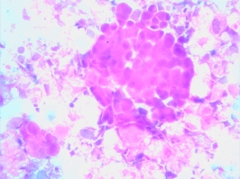
Sarcoma 130412.01With the profusion of genomic analyses available today at virtually every medical center, it came as no surprise that both patients had undergone genetic profiling. What struck me were the results. The young woman had “no measurable genetic aberrancies” from a panoply of 370 cancer-causing exomes, while the young man’s tumor revealed no somatic mutations and only two germ-line SNV’s (single nucleotide variants) from a 50 gene NextGen sequence, neither of which had any clinical or therapeutic significance.

What are we to make of these findings? By conventional wisdom, cancer is a genetic disease. Yet, neither of these patients carried detectable “driver” mutations. Are we to conclude that the tumors that invaded the cervical vertebra of the young woman, requiring an emergency spinal fusion, or the large mass in the lung of the young man are not “cancers”? It would seem that if we apply contemporary dogma, these patients do not have a cancer at all. But nothing could be further from the truth.

Cancer as a disease is not a genomic phenomenon. It is a phenotypic one. As such, it is extremely likely that these patients’ tumors are successfully exploiting normal genes in abnormal ways. The small interfering RNAs or methylations or acetylation or non-coding DNA’s that conspired to create these monstrous problems are too deeply encrypted to be easily deciphered by our DNA methodologies. These changes are effectively gumming up the works of the cancer cell’s biology without leaving a fingerprint.  Slide Detail-small

I have long recognized that cellular studies like the EVA-PCD platform provide the answers, through functional profiling, that genetic analyses can only hope to detect. The assay did identify drugs active in these patients’ tumor, which will offer meaningful benefit, despite the utter lack of genetic targets. Once again, we are educated by cellular biology in the absence of genomic insights. This leaves us with a question however – is cancer a genetic disease?
Is There a Role for PI3k Inhibitors in Breast Cancer? Maybe.

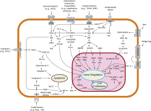
Over the past decades oncologists have learned that cancer is driven by circuits known as signal transduction pathways. Signal_transduction_pathways.svgThe first breakthroughs were in chronic myelogenous leukemia (CML) where a short circuit in the gene as c-Abl caused the overgrowth of malignant blasts. The development of Imatinib (Gleevec) a c-Abl inhibitor yielded brilliant responses and durable remissions with a pill a day.

The next breakthrough came with the epidermal growth factor pathway and the development of Gefitinib (Iressa) and shortly thereafter Erlotinib (Tarceva). Good responses in lung cancers, many durable were observed and the field of targeted therapy seemed to be upon us.

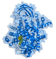
220px-PI3kinaseAmong the other signal pathways that captured the imagination of the pharmaceutical industry as a potential target was phospho-inositol-kinase (PI3K). Following experimental work by Lew Cantley, PhD, who first described this pathway in 1992, more than a dozen small molecules were developed to inhibit this cell signal system.

The PI3K pathway is important for cell survival and regulates metabolic activities like glucose uptake and protein synthesis. It is associated with insulin signaling and many bio-energetic phenomena. The earliest inhibitors functioned downstream at a protein known as mTOR, and two have been approved for breast, neuroendocrine and kidney cancers. Based on these early successes, PI3K, which functions upstream and seemed to have much broader appeal, became a favored target for developmental clinical trials.

The San Antonio Breast Cancer Symposium is one of the most important forums for breast cancer research. The December 2014 meeting featured a study that combined one of the most potent PI3K inhibitors, known as Pictilisib, with a standard anti-estrogen drug, Fulvestrant, in women with recurrent breast cancer. The FERGI Trial only included ER positive patients who had failed prior treatment with an aromatase inhibitor (Aromasin, Arimidex or Femara). The patients were randomized to receive the ER blocker Fulvestrant with or without Pictilisib.

With seventeen months of follow-up there was some improvement in time to progressive disease, but this was not large enough to achieve significance and the benefit remains unproven. A subset analysis did find that for patients who were both ER (+) and PR (+) a significant improvement did occur. The ER & PR (+) patients benefitted for 7.4 months on the combination while those on single agent Fulvestrant for only for 3.7 months.

The FERGI trial is more interesting for what it did not show. And that is that patients who carried the PI3K mutation, the target of Pictilisib, did not do better than those without mutation (known as wild type). To the dismay of those who tout the use of genomic biomarkers like PI3K mutation for patient drug selection, the stunning failure to identify responders at a genetic level should send a chill down the spine of every investor who has lavished money upon the current generation of genetic testing companies.

It should also raise concerns for the new federal programs that have designated hundreds of millions of dollars on the new “Personalized Cancer Therapy Initiatives” based entirely on genomic analyses. The contemporary concept of personalized cancer care is explicitly predicated upon the belief that genomic patient selection will improve response rates, reduce costs and limit exposure to toxic drugs in patients unlikely to respond.

This unanticipated failure is only the most recent reminder that genomic analyses can only suggest the likelihood of response and are not determinants of clinical outcome even in the most enriched and carefully selected individuals. It is evident from these findings that PI3K mutation alone doesn’t define the many bioenergetic pathways associated with the phenotype. This strongly supports phenotypic analyses like EVA-PCD as better predictors of response to agents of this type, as we have shown in preclinical and clinical analyses.

Bevacizumab In Colon Cancer – “A Shot Across The Bowel”

Colon2 130320.01 lo resAn E-Publication article in the February Journal of Clinical Oncology analyzes the cost efficacy of Bevacizumab for colon cancer. Bevacizumab, sold commercially as Avastin, has become a standard in the treatment of patients with advanced colorectal cancer. Indeed, Bevacizumab plus FOLFOX or FOLFIRI, are supported by NCCN guidelines and patients who receive one of these regimens are usually switched to the other at progression.

A Markov computer model explored the cost and efficacy of Bevacizumab in the first and second line setting using a well-established metric known as a Quality-Adjusted Life Year (QALY). In today’s dollars $100,000 per QALY is considered a threshold for utility of any treatment. To put this bluntly, the medical system values a year of yavastinour life at $100,000. The authors confirmed that Bevacizumab prolongs survival but that it does so at significantly increased costs. By their most optimistic projections, Bevacizumab + FOLFOX come in at more than $200,000 per QALY. Similar results were reported for Canadian, British and Japanese costs. Though more favorable, the results with FOLFIRI + Bevacizumab still came in above the $100,000 threshold.

No one doubts that Bevacizumab provides improved outcomes. It’s the incremental costs that remain an issue. Society is now confronting an era where the majority of new cancer agents come in at a cost in excess of $10,000 per month. Where and how will we draw the line that designates some treatments unaffordable? On the one hand, clinical therapies could be made available only to the “highest bidder.” However, this is contrary to the western societal ethic that holds that medical care should be available to all regardless of ability to pay. Alternatively, increasingly narrow definitions could be applied to new drugs making these treatments available to a shrinking minority of those who might actually benefit; a form of “evidence-based” rationing. A much more appealing option would be to apply validated drug predication assays for the intelligent selection of treatment candidates.
Avastin-MOA-Overview
In support of the latter, the authors state, “Bevacizumab potentially could be improved with the use of an effective biomarker to select patients most likely to benefit.” This is something that genomic (DNA) profiling has long sought to achieve but, so far, has been unable to do. This conceptual approach however is demonstrably more attractive in that all patients have equal access, futile care is avoided and the costs saved would immediately provide highly favorable QALY’s as the percentage of responders improved.

Similar to the recent reports from the National Health Service of England, the American public now confronts the challenge of meeting the needs of a growing population of cancer patients at ever-higher costs. It is only a matter of time before these same metrics described for colon cancer are applied to lung, ovarian and other cancers for which Avastin is currently approved.

At what point will the American medical system recognize the need for validated predictive platforms, like EVA-PCD analyses, that have the proven capacity to save both money and lives? We can only wonder.

Cancer Patients Need Answers Now!

I read a sad editorial in the Los Angeles Times written by Laurie Becklund, former LA Times journalist. It is, in essence, a self-written obituary as the patient describes her saga beginning almost 19 years earlier, when she detected a lump in her breast. With stage I breast cancer she underwent standard therapy and remained well for 13 years until recurrence was heralded by disease in bone, liver, lung and brain. Given a dire prognosis she became a self-made expert, conducting research, attending conferences, and joining on-line forums under the name “Won’t Die of Ignorance.” Despite her heroic effort Ms. Becklund succumbed to her illness on February 8. She was 66.

Ms. Becklunla-laurie-becklund-cropp-jpg-20150209d experienced the anguish that every patient feels when his or her own individual and highly personal needs simply aren’t being addressed. She opines that entities like the Susan G. Komen Fund, which has raised over $2.5 billion in the last 20 years, “channels only a fraction of those funds into research or assistance to help those who are already seriously sick.” She continues, “We need people, patients, doctors, scientists, politicians, industry and families to make a fresh start.” Her frustration is palpable as she states her outcome seemed to be based on the roll of the dice, like playing “Chutes and Ladders.”

The author’s plight is shared by the millions of patients who are confronting advanced cancers. They are not interested in “why” or “how” their cancers came to be. They can no longer benefit from early detection or cancer awareness campaigns. They need practical, actionable, clinical answers today.

Ms. Becklund’s commentary resonates with me and with everyone who has cOutliving Cancerancer or knows someone who does. As an oncology fellow at Georgetown, I found myself losing patient after patient to toxic and largely ineffective treatments, all despite my best efforts. I described this in my book “Outliving Cancer.” It was then that I decided that I would dedicate myself to meeting the individual needs of each of my patients and I have used a laboratory platform (EVA-PCD) to do so. I have encountered surprising resistance from clinicians and researchers who seem to prefer the glacial pace of incremental advancement found in population studies over individual solutions found in the study of each patient’s unique biology. Ms. Becklund correctly points out that every treatment must meet each individual’s need.

The role of the scientist is to answer a question (treatment A vs. treatment B) while that of the clinical physician must be to save a life. Every patient is an experiment in real time. It may well be that no two cancer patients are the same. Indeed, the complexity of carcinogenesis makes it very possible that every patient’s cancer is an entirely new disease, never before encountered. Although cancers may look alike, they may be biologically quite distinct. Meaningful advances in cancer will only occur when we learn to apply all available technologies to treat patients as the individuals that they are. Let us hope that Ms. Becklund’ s final essay does not fall upon deaf ears.

A New Use for One of the Oldest “New” Drugs

With the profusion of new targeted agents entering the clinical arena, a report from the American Society of Hematology bears consideration.

The trial known as the SORAML trial enrolled 276 patients with newly diagnosed acute myelogenous leukemia. The patients were between the ages of 18 and 60. All patients received a standard chemotherapy regimen. The patients were then randomized to receive Sorafenib or placebo. Patients on the Sorafenib arm then remained on a maintenance therapy for twelve months.

While the achievement of complete remission was almost identical between the two arms at 59% and 60%, the event free survival demonstrably favored the Sorafenib group at 20.5 months versus 9.2 months. At three years of follow-up 40% of the Sorafenib group were well with only 22% of the placebo group still in remission. This corresponds to a three-year relapse free survival of 38% for placebo and 56% for Sorafenib (P=0.017).

The results are of interest on several levels.
1.    Sorafenib a multitargeted tyrosine kinase inhibitor was approved in December 2005 for the treatment of renal cell carcinoma. This makes Sorafenib one of the first targeted agents to achieve FDA approval.

2.     Sorafenib has many modes of action and it is not entirely clear which of its functions were responsible for the superior survival in this AML study.

3.    Sorafenib’s approval reflects a rather convoluted and interesting history. When first developed the drug was designed to target the oncogene B-Raf. As a result the drug was introduced into early clinical trials for the treatment of advanced melanoma, a disease known to be associated with B-Raf mutation. As the drug proved ineffective, it appeared unlikely to gain FDA approval. That is, until it showed cross reactivity with VEGF pathway associated with tumor cell vascularity. A successful trial published in the New England Journal of Medicine then led to the approval.

Now, nine years later this old new drug has gained new life. This time in acute myelogenous leukemia.

The term “dirty drug” refers to agents that target many kinases at the same time. Sorafenib is an example of a “dirty drug.” However it is Sorafenib’s “dirty drug” quality that led first to its approval and most likely now leads to its application in AML. This reflects the fact that Sorafenib may be inhibiting B-Raf signaling associated with the common mutation in Ras upstream of B-Raf or it may reflect Flt3 a secondary activity associated with Sorafenib.

Indeed B-Raf and Flt3 may not be upregulated in every patient, but could serve a function of permissive activity granting an additional survival signal to the AML cells as they go through induction therapy. These subtleties of drug effect may escape genomic analysis as the true “target” may not be mutated, upregulated or amplified. No doubt the investigators in this study will conduct gene sequencing to determine whether there is a driver mutation associated with the advantage reported in this clinical study. What will be intriguing is to determine whether that advantage is an abnormal gene functioning within these cancerous cells or possibly a normal gene functioning abnormally in these cancer cells. More to come.

Genomic Profiling for Lung Cancer: the Good, the Bad and the Ugly

Genomic profiling has gained popularity in medical oncology. Using NextGen platforms, protein coding regions of human tumors known as exomes can be examined for mutations, amplifications, deletions, splice variants and SNPs. In select tumors the results can be extremely helpful. Among the best examples are adenocarcinomas of the lung where EGFr, ALK and ROS-1 mutations, deletions and/or re-arrangements identified by DNA analysis can guide the selection of “targeted agents” like Erlotinib and Crizotinib.

An article published in May 2014 issue of JAMA reported results using probes for 10 “oncogenic driver” mutations in lung cancer patients. They screened for at least one gene in 1,007 patients and all 10 genes in 733. The most common was k-ras at 25%, followed by EGFR in 17% and ALK in 8%. The incidence then fell off with other EGFr mutations in 4%, B-raf mutations in 2%, with the remaining mutations each found in less than 1%.

Median survival at 3.5 vs 2.4 years was improved for patients who received treatments guided by the findings (Kris MG et al, Using multiplex assays of oncogenic drivers in lung cancers to select targeted drugs. JAMA, May 2014). Do these results indicate that genomic analyses should be used for treatment selection in all patients? Yes and no.

Noteworthy is the fact that 28% of the patients had driver mutations in one of three genes, EGFr, HER2 or ALK. All three of these mutations have commercially available chemotherapeutic agents in the form of Erlotinib, Afatinib and Crizotinib. Response rates of 50% or higher, with many patients enjoying durable benefits have been observed. Furthermore, patients with EGFr mutations are often younger, female and non-smokers whose tumors often respond better to both targeted and non-targeted therapies. These factors would explain in part the good survival numbers reported in the JAMA article. Today, a large number of commercial laboratories offer these tests as part of standard panels. And, like k-ras mutations in colon cancer or BCR-abl in CML (the target of Gleevec), the arguments in favor of the use of these analyses is strong.

Non-small cell lung cancer

Non-small cell lung cancer

But what of the NSCLC patients for whom no clear identifiable driver can be found? What of the 25% with k-ras mutations for whom no drug exists? What of those with complex mutational findings? And finally what of those patients whose tumors are driven by normal genes functioning abnormally? In these patients no mutations exists at all. How best do we manage these patients?

I was reminded of this question as I reviewed a genomic analysis reported to one of my colleagues. He had submitted a tissue block to an east coast commercial lab when one of his lung cancer patients relapsed. The results revealed mutations in EGFr L858R & T790M, ERBB4, HGF, JAK2, PTEN, STK11, CCNE1, CDKN2A/B, MYC, MLL2 W2006, NFKB1A, and NKX2-1. With a tumor literally bristling with potential targets, what is a clinician to do? How do we take over a dozen genetically identified targets and turn them into effective treatment strategies? In this instance, too much information can be every bit as paralyzing as too little.

Our preferred approach is to examine the small molecule inhibitors that target each of the identified aberrancies in our laboratory platform. We prefer to drill down to the next level of certainty e.g. cellular function. After all, the presence of a target does not a response make.

In this patient I would conduct a biopsy. This would enable us to examine the drugs and combinations that are active against the targets. A “hit” by the EVA-PCD assay would then isolate the “drivers” from the “passengers” and enable the clinician to intelligently select effective treatments. Combining genomic analyses with functional profiling (phenotypic analyses) provides the opportunity to turn speculative observations into actionable events.

This is the essence of Rational Therapeutics.

New Diagnostic Test for the Early Detection of Lung Cancer

I was invited to discuss a new diagnostic test for the early detection of lung cancer by Gerri Willis of Fox Business News’ Willis Report.
40-110-000-web
An Italian clinical study presented at the September 2014 European Respiratory Society described 82 patients with abnormal chest x-rays. Patients breathed into a machine that measured the temperature of the exhaled air. Forty of the patients ultimately proved to have cancer and 42 did not, as confirmed by subsequent biopsy. They found a correlation between the temperature of the exhaled breath and presence of lung cancer. They also found that long term smokers had higher breath temperatures, as did those with higher stage disease.

For a variety of reasons, a test as simple as breath temperature seems unlikely to be highly specific. After all, the temperature of the exhaled breath could reflect infection, inflammation, or even activity level, as vigorous exercise can raise the body’s core temperature. Nonetheless, the fact that there is any correlation at all is of interest.

PET scan lung cancerWhat might underlie these findings? Accepting the shortfalls of this small study, it is an interesting point of discussion. First, cancer is a hyper metabolic state. Cancers consume increased quantities of glucose, proteins, and lipids. PET scans measure these phenomena every day. Second, cancer is associated with hyper vascularity. Up-regulation of VEGF could cause hyperemia (increased capillary blood flow) in the airways of lung cancer patients, resulting in the finding. Finally, cancer, in and of itself, is an inflammatory state. Inflammation reflects increased metabolic activity that could manifest as a whole body change in basal temperature.

Lung cancer is the leading cause of cancer death in the US, constituting 27% of all cancer deaths. Despite the over 224,000 new diagnoses and 160,000 deaths, the five-year survival for lung cancer today at 17% has not changed in several decades. Nonetheless patients who are detected early (Stage I) have a greater than 50% five-year survival.

We know from the National Lung Cancer Screening Trial published in 2010, that early detection by CT scans can reduce mortality from this disease by 20%. In the cancer literature, that is huge. The problem is that screening CTs are comparatively expensive, inconvenient, expose patients to radiation and are themselves fraught with false positives and false negatives. Furthermore, it is estimated that that broad application of spiral CT’s could cost over $9 billion a year. Thus, simple, non-invasive screening techniques are sorely needed.

The use of exhaled breath to diagnose cancers has been under in development for decades. Recently, investigators from The Cleveland Clinic and others from Israel have reported good results with a microchip that measures the concentration of volatile organic compounds in the breath and provides a colorimetric score. With several hundred patients the receiver-operating curves (ROC, a technique that gauges the sensitivity and specificity of a test) in the range of 0.85 (1.0 is perfect) are quite favorable. Although these techniques have not yet gained broad application, they are extremely interesting from the standpoint of what it is they are actually measuring.

For decades, the principal focus of scientific exploration in cancer has been genomic. Investigators at Boston University and others at MD Anderson in Texas have used genomic and methylation status of oro-and naso-pharyngeal swabs to identify the earliest hallmarks of malignant transformation. To the contrary, the breath tests described above measure phenomena that fall more in the realm of metabolomics. After all, these are measures of cellular biochemical reactions and identify the transformed state at a metabolic level.

Though still in its infancy, metabolomics reflects the most appealing of all cancer analyses. Examining cancer for what it is, rather than how it came to be, uses biochemistry, enzymology and quantitative analyses. These profile the tumor at the level of cellular function. Like the platforms that I utilize (EVA-PCD), these metabolic analyses examine the tumor phenotype.

I applaud these Italian investigators for using a functional approach to cancer biology. This is a highly productive direction and fertile ground for future research. Will breath temperature measurement prove sensitive and specific enough to diagnose cancer at early stage? It is much too early to say, but at least for now, I wouldn’t hold my breath.

Toward A 100% Response Rate in Human Cancer

Oncologists confront numerous hurdles as they attempt to apply the new cancer prognostic and predictive tests. Among them are the complexities of gene arrays that introduce practicing physicians to an entirely new lexicon of terms like “splice variant, gene-rearrangement, amplification and SNP.”

Althougcancer for dummiesh these phrases may roll of the tongue of the average molecular biologists (mostly PhDs), they are foreign and opaque to the average oncologist (mostly MDs). To address this communication shortfall laboratory service providers provide written addenda (some quite verbose) to clarify and illuminate the material. Some institutions have taken to convening “molecular tumor boards” where physicians most adept at genomics serve as “translators.” Increasingly, organizations like ASCO offer symposia on modern gene science to the rank and file, a sort of Cancer Genomics for Dummies. If we continue down this path, oncologists may soon know more but understand less than any other medical sub-specialists.

However well intended these educational efforts may be, none of them are prepared to address the more fundamental question: How well do genomic profiles actually predict response? This broader issue lays bare our tendency to confuse data with results and big data with big results. To wit, we must remember that our DNA, originally provided to each of us in the form of a single cell (the fertilized ovum) carries all of the genetic information that makes us, us. From the hair follicles on our heads to the acid secreting cells in our stomach, every cell in our body carries exactly the same genetic data neatly scripted onto our nuclear hard-drives.
Forest
What makes this all work, however, isn’t the DNA on the hard drive, but instead the software that judiciously extracts exactly what it needs, exactly when it needs it. It’s this next level of complexity that makes us who we are. While it is true that you can’t grow hair or secrete stomach acid without the requisite DNA, simply having that DNA does not mean you will grow hair or make acid. Our growing reliance upon informatics has created a “forest for the trees” scenario, focusing our gaze upon nearby details at the expense of larger trends and insights.

What is desperately needed is a better approximation of the next level of complexity. In biology that moves us from the genotype (informatics) to the phenotype (function). To achieve this, our group now regularly combines genomic, transcriptomic or proteomic information with functional analyses. This enables us to interrogate whether the presence or absence of a gene, transcript or protein will actually confer that behavior or response at the system level.

I firmly believe that the future of cancer therapeutics will combine genomic, transcriptomic and/or proteomic analyses with functional (phenotypic) analyses.

Recent experiences come to mind. A charming patient in her 50s underwent a genomic analysis that identified a PI3K mutation. She sought an opinion. We conducted an EVA-PCD assay on biopsied tissue that confirmed sensitivity to the drugs that target PI3K. Armed with this information, we administered Everolimus at a fraction of the normal dose. The response was prompt and dramatic with resolution of liver function abnormalities, normalization of her performance status and a quick return to normal activities. A related case occurred in a young man with metastatic colorectal cancer. He had received conventional chemotherapies but at approximately two years out, his disease again began to progress.

A biopsy revealed that despite prior exposure to Cetuximab (the antibody against EGFR) there was persistent activity for the small molecule inhibitor, Erlotinib. Consistent with prior work that we had reported years earlier, we combined Cetuximab with Erlotinib, and the patient responded immediately.

Each of these patients reflects the intelligent application of available technologies. Rather than treat individuals based on the presence of a target, we can now treat based on the presence of a response. The identification of targets and confirmation of response has the potential to achieve ever higher levels of clinical benefit. It may ultimately be possible to find effective treatments for every patient if we employ multi-dimensional analyses that incorporate the results of both genomic and phenotypic platforms.

With an EVA-PCD Assay, It Can Be That Simple

Shortly after I left the university and joined a medical oncology group, one of the junior members of the practice asked if I would cover for him during his summer vacation. Among the patients he signed over to me was a gentleman in his 60s with what he described as “end-stage” chronic lymphocytic leukemia (CLL). As the patient had already received the standard therapies, second line regimens and experimental drugs available at the time, the physician had run out of options. My charge was to keep him comfortable. I asked if it would be all right for me to study his cells in my lab and the doctor agreed.

CLL 130611.06I met the patient the next day. He was a very pleasant tall, slender black man lying in bed. He had lost a great deal of weight making the already enlarged lymph nodes in his neck appear that much more prominent. As I was engaged in the study of CLL as my principal tumor model, I asked if I might examine his circulating CLL cells as part of our IRB-approved protocol. He graciously obliged and I obtained a few ccs of blood. We were deeply ensconced in tumor biology analyses and his cells were used to explore membrane potentials, DNA degradation and glutathione metabolism as correlates with drug response profiles by EVA-PCD analysis. A large number of those studies have since been published.

What struck me about the patient’s EVA-PCD profile was the exquisite sensitivity to corticosteroids. Corticosteroids in the form of prednisone, Medrol, Solu-Medrol and Decadron are the mainstays of therapy for lymphoid malignancies like CLL. Everyone receives them. Indeed this patient had received them repeatedly including his first line chlorambucil plus prednisone, his second-line CHOP and his third line ESHAP. It was only after he had failed all of these increasingly intensive regimens that he finally moved on to an experimental agent, homoharringtonine, a drug that finally received FDA approval in 2012, after almost 40 years of clinical development. Unfortunately for him homoharringtonine did not work and it seemed we were well beyond conventional therapies, or were we?

I pondered the corticosteroid sensitivity finding and decided to start the patient on oral prednisone. It would be another two weeks before his physician returned and there really weren’t many options. The patient responded overnight. The lymph nodes melted away. The spleen diminished. He began to eat and gained weight. Within a few days he felt well enough to go home. I discharged the patient and remember writing his prednisone prescription, 40 mg by mouth each morning.

A week later, my colleague returned from his retreat in the Adirondacks. He inquired about his patients and surmised that this gentleman, no longer in the hospital, had died. I explained that he had been discharged.

“Discharged . . . how?” he asked. I described the findings of our EVA-PCD study, the sensitivity to steroids and the patient’s miraculous clinical response to this, the simplest of all possible treatments. The physician then turned to me and said “Prednisone . . . hmmm . . . I could have done that.”

I am reminded of this story almost daily. It is emblematic of our work and of those who choose not to use it. Good outcomes in cancer do not occur by chance. They also do not require blockbuster new drugs or brilliant doctors. They require individualized attention to the needs of each patient.

A recurring theme, exemplified by this patient among others, is that cancer cells can only defend themselves in a limited number of ways. Once a selection pressure, in a Darwinian sense, is removed (e.g. corticosteroids were not used during the homoharringtonine treatments) the surviving cells, sensitive to steroids, re-emerge to be identified and captured in our laboratory platform.

It is remarkable how often heavily pretreated patients with ovarian cancer are found sensitive to Taxol after they had received it years earlier, but not since; or breast cancer patients who fail every new agent only to prove responsive to CMF, the earliest of all of the breast cancer drug combinations developed in the 1970s. Our job as oncologists is to find those chinks in armor of cancer cells and exploit them. The EVA-PCD platform, in the eyes of some, may not be groundbreaking . . . it just happens to work!


The Changing Landscape in Non-small Cell Lung Cancer (NSCLC)

In October 2012, we published a study of patients with metastatic NSCLC whose treatment was guided by EVA-PCD laboratory analysis. The trial selected drugs from FDA approved, compendium listed chemotherapies and every patient underwent a surgical biopsy under an IRB-approved protocol to provide tissue for analysis.

The EVA-PCD patients achieved an objective response rate of 64.5 percent (2-fold higher than national average, P < 0.0015) and median overall survival of 21.3 months (nearly 2-fold longer than the national average of 12.5 months).

Non-small cell lung cancer

Non-small cell lung cancer

The concept of conducting biopsies in patients with metastatic NSCLC was not only novel in 2004, it was downright heretical. Physicians argued forcefully that surgical procedures should not be undertaken in metastatic disease fearing risks and morbidity. Other physicians were convinced that drug selection could not possibly improve outcomes over those achieved with well-established NCCN guidelines. One oncologist went so far as to demand a formal inquiry. When the hospital was forced to convene an investigation, it was the co-investigators on the IRB approved protocol and the successfully treated patients who ultimately rebuffed this physician’s attempt to stifle our work.

With the publication of our statistically superior results and many of our patients surviving more than 5 years, we felt vindicated but remain a bit battle scarred.

I was amused when one of my study co-authors (RS) recently forwarded a paper authored at the University of California at Davis about surgical biopsies and tumor molecular profiling published by The Journal of Thoracic and Cardiovascular Surgery. This single institution study of twenty-five patients with metastatic NSCLC reported their experience-taking patients with metastatic disease to surgical biopsy for the express purpose of selecting therapy. Sixty four percent were video assisted thoracic (VATS) wedge biopsies, 16 percent pleural biopsies, 8 percent mediastinoscopies, 12 percent supraclavicular biopsies and 8 percent rib/chest wall resections. Tissues were submitted to a commercial laboratory in Los Angeles for genomic profiling.

The authors enthusiastically described their success conducting surgical procedures to procure tissue for laboratory analysis. Gone was the anxiety surrounding the risk of surgical morbidity. Gone were the concerns regarding departure from “standard” treatment. In their place were compelling arguments that recapitulated the very points that we had articulated ten years earlier in our protocol study. While the platforms may differ, the intent, purpose and surgical techniques applied for tissue procurement were exactly the same.

What the Cooke study did not describe was the response rate for patients who received “directed therapy.” Instead they provide the percent of patients with “potentially targetable” findings (76 percent) and the percent that had a “change in strategy” (56 percent) as well as those that qualified for therapeutic trials (40 percent). Though, laudable, changing strategies and qualifying for studies does not equal clinical responsiveness. One need only examine the number of people who are “potential winners” at Black Jack or those who “change their strategies” (by changing tables/dealers for example) or, for that matter, those who qualify for “high roller status” to understand the limited practical utility of these characterizations.

Nonetheless, the publication of this study from UC Davis provides a landmark in personalized NSCLC care. It is no longer possible for oncologists to decry the use of surgical biopsies for the identification of active treatments.

As none of the patients in this study signed informed consents for biopsy, we can only conclude that the most august institutions in the US now view such procedures as appropriate for the greater good of their patients. Thus, we are witness to the establishment of a new paradigm in cancer medicine. Surgical biopsies in the service of better treatment are warranted, supported and recommended. Whatever platform, functional or genomic, patient-directed therapy is the new normal and the landscape of lung cancer management has changed for the better.