Is Cancer a Genetic Disease?

I recently had the opportunity to meet two charming young patients: One, a 32-year-old female with an extremely rare malignancy that arose in her kidney and the other a 33-year-old gentleman with widely metastatic sarcoma.

Both patients had obtained expert opinions from renowned cancer specialists and both had undergone aggressive multi-modality therapies including chemotherapy, radiation and surgery. Although they suffered significant toxicities, both of their diseases had progressed unabated. Each arrived at my laboratory seeking assistance for the selection of effective treatment.
Sarcoma 130412.01With the profusion of genomic analyses available today at virtually every medical center, it came as no surprise that both patients had undergone genetic profiling. What struck me were the results. The young woman had “no measurable genetic aberrancies” from a panoply of 370 cancer-causing exomes, while the young man’s tumor revealed no somatic mutations and only two germ-line SNV’s (single nucleotide variants) from a 50 gene NextGen sequence, neither of which had any clinical or therapeutic significance.

What are we to make of these findings? By conventional wisdom, cancer is a genetic disease. Yet, neither of these patients carried detectable “driver” mutations. Are we to conclude that the tumors that invaded the cervical vertebra of the young woman, requiring an emergency spinal fusion, or the large mass in the lung of the young man are not “cancers”? It would seem that if we apply contemporary dogma, these patients do not have a cancer at all. But nothing could be further from the truth.

Cancer as a disease is not a genomic phenomenon. It is a phenotypic one. As such, it is extremely likely that these patients’ tumors are successfully exploiting normal genes in abnormal ways. The small interfering RNAs or methylations or acetylation or non-coding DNA’s that conspired to create these monstrous problems are too deeply encrypted to be easily deciphered by our DNA methodologies. These changes are effectively gumming up the works of the cancer cell’s biology without leaving a fingerprint.  Slide Detail-small

I have long recognized that cellular studies like the EVA-PCD platform provide the answers, through functional profiling, that genetic analyses can only hope to detect. The assay did identify drugs active in these patients’ tumor, which will offer meaningful benefit, despite the utter lack of genetic targets. Once again, we are educated by cellular biology in the absence of genomic insights. This leaves us with a question however – is cancer a genetic disease?

Investigators in Boston Re-Invent the Wheel

A report published in Cell from Dana-Farber Cancer Institute describes a technique to measure drug cov150hinduced cell death in cell lines and human cancer cells. The method “Dynamic BH3 profiling” uses an oligopeptidic BIM to gauge the degree to which cancer cells are “primed” to die following exposure to drugs and signal transduction inhibitors. The results are provocative and suggest that in cell lines and some human primary tissues, the method may select for sensitivity and resistance.

We applaud these investigators’ recognition of the importance of phenotypic measures in drug discovery and drug selection and agree with the points that they raise regarding the superiority of functional platforms over static (omic) measures performed on paraffin fixed tissues. It is heartening that scientists from so august an institution as Dana-Farber should come to the same understanding of human cancer biology that many dedicated researchers had pioneered over the preceding five decades.

Several points bear consideration. The first, as these investigators so correctly point out: “DBP should only be predictive if the mitochondrial apoptosis pathway is being engaged.” This underscores the limitation of this methodology in that it only measures one form of programmed cell death – apoptosis. It well known that apoptosis is but one of many pathways of programmed cell death, which include necroptosis, autophagy and others.

While leukemias are highly apoptosis driven, the same cannot so easily be said of many solid tumors like colon, pancreas and lung. That is, apoptosis may be a great predictor of response except when it is not. The limited results with ovarian cancers (also apoptosis driven) are far from definitive and may better reflect unique features of epithelial ovarian cancers among solid tumors than the broad generalizability of the technique.

A second point is that these “single cell suspensions” do not recreate the microenvironment of human tumors replete with stroma, vasculature, effector immune cells and cytokines. As Rakesh Jain, a member of the same faculty, and others have so eloquently shown, cancer is not a cell but a system. Gauging the system by only one component may grossly underestimate the systems’ complexity, bringing to mind the allegory of elephant and the blind man. Continuing this line of reasoning, how might these investigators apply their technique to newer classes of drugs that influence vasculature, fibroblasts or stroma as their principal modes of action? It is now recognized that micro environmental factors may contribute greatly to cell survival in many solid tumors. Assay systems must be capable of capturing human tumors in their “native state” to accurately measure these complex contributions.

Thirdly, the ROC analyses consistently show that this 16-hour endpoint highly correlates with 72- and 96-hour measures of cell death. The authors state, “that there is a significant correlation between ∆% priming and ∆% cell death” and return to this finding repeatedly. Given that existing short term (72 – 96 hour) assays that measure global drug induced cell death (apoptotic and non-apoptotic) in human tumor primary cultures have already established high degrees of predictive validity with an ROC of 0.89, a 2.04 fold higher objective response rate (p =0.0015) and a 1.44 fold higher one-year survival (p = 0.02) are we to assume that the key contribution of this technique is 56 hour time advantage? If so, is this of any clinical relevance? The report further notes that 7/24 (29%) of ovarian cancer and 5/30 (16%) CML samples could not be evaluated, rendering the efficiency of this platform demonstrably lower than that of many existing techniques that provide actionable results in over 90% of samples.

Most concerning however, is the authors’ lack of recognition of the seminal influence of previous investigators in this arena. One is left with the impression that this entire field of investigation began in 2008. It may be instructive for these researchers to read the first paper of this type in the literature published in in the JNCI in 1954 by Black and Spear. They might also benefit by examining the contributions of dedicated scientists like Larry Weisenthal, Andrew Bosanquet and Ian Cree, all of whom published similar studies with similar predictive validities many years earlier.

If this paper serves to finally alert the academic community of the importance of human tumor primary culture analyses for drug discovery and individual patient drug selection then it will have served an important purpose for a field that has been grossly underappreciated and underutilized for decades. Mankind’s earliest use of the wheel dates to Mesopotamia in 3500 BC. No one today would argue with the utility of this tool. Claiming to have invented it anew however is something quite different.

In Cancer Research: An Awakening?

In 2005, as the Iraq War reached a low point with casualties mounting and public support dwindling, Sunni tribesman in the Anbar Province arose to confront the enemy. Joining together as an ad hoc army these fighters turned the tide of the war and achieved victories in the face of what had appeared at the time, to be overwhelming odds.

I am reminded of this by an article in The Wall Street Journal by Peter Huber and Paul Howard of the Manhattan Institute that examined the bureaucracy of drug development. It raised the question: Are new cancer treatments failures or is the process by which they are approved a failure? They describe “exceptional responders” defined as patients who show unexpected benefits from drug treatments. Using molecular profiles, they opine, scientists will unravel the mysteries of these individuals and usher in an era of personalized medicine. Thus, rigid protocols that use drugs based upon tumor type e.g. lung vs. colon fail because they do not incorporate the features that make each patient unique – an awakening.

The example cited is from Memorial Sloan-Kettering where a patient with bladder cancer had an unexpected response to the drug Everolimus (approved for kidney cancer). Subsequent deep sequencing identified a genetic signature associated with sensitivity to this drug. While it is a nice story, I already knew it very well because it had been repeated many times before and would in the past have been dismissed as an “anecdote.” It is precisely because of its rarity that it has been repeated so many times.

The WSJ analysis strikes a familiar chord. For decades, we have decried the failure of rigid clinical trials that underestimate a patient’s unique biology yet cost millions, even billions of dollars, while denying worthy candidates new treatments under stultifying disease-specific designs.

Well Tray Closeup2 smallWe pioneered phenotypic (functional) analyses (the EVA-PCD platform) to examine whole cell models as we explored drug response profiles, novel combinations and new targets. It is regrettable that these WSJ authors, having raised such important issues, then stumble into the same tantalizing trap of molecular diagnostics, and call for bigger, better, faster genomic analyses.

Cancer patients need to receive treatments that work. They do not particularly care why or how they work, just that they work. These authors seem to perpetuate the myth that we must first understand why a patient responds before we can treat them. Nothing could be further from the truth.

Alexander Fleming knew little about bacterial cell wall physiology when he discovered penicillin in 1928, and William Withering knew nothing about the role of muscle enzymes in congestive heart failure when he discovered digoxin extracts in 1785. Would anyone argue that we should have waited decades, even centuries to apply manifestly effective therapies to patients because we did not have the “genes sequenced?’

We may be witness to an awakening in cancer drug development. It may be that a new understanding of individualized patient response will someday provide better outcomes, but platforms with the proven capacity to connect patients to available treatments should be promoted and applied today.

Genomic Profiling for Lung Cancer: the Good, the Bad and the Ugly

Genomic profiling has gained popularity in medical oncology. Using NextGen platforms, protein coding regions of human tumors known as exomes can be examined for mutations, amplifications, deletions, splice variants and SNPs. In select tumors the results can be extremely helpful. Among the best examples are adenocarcinomas of the lung where EGFr, ALK and ROS-1 mutations, deletions and/or re-arrangements identified by DNA analysis can guide the selection of “targeted agents” like Erlotinib and Crizotinib.

An article published in May 2014 issue of JAMA reported results using probes for 10 “oncogenic driver” mutations in lung cancer patients. They screened for at least one gene in 1,007 patients and all 10 genes in 733. The most common was k-ras at 25%, followed by EGFR in 17% and ALK in 8%. The incidence then fell off with other EGFr mutations in 4%, B-raf mutations in 2%, with the remaining mutations each found in less than 1%.

Median survival at 3.5 vs 2.4 years was improved for patients who received treatments guided by the findings (Kris MG et al, Using multiplex assays of oncogenic drivers in lung cancers to select targeted drugs. JAMA, May 2014). Do these results indicate that genomic analyses should be used for treatment selection in all patients? Yes and no.

Noteworthy is the fact that 28% of the patients had driver mutations in one of three genes, EGFr, HER2 or ALK. All three of these mutations have commercially available chemotherapeutic agents in the form of Erlotinib, Afatinib and Crizotinib. Response rates of 50% or higher, with many patients enjoying durable benefits have been observed. Furthermore, patients with EGFr mutations are often younger, female and non-smokers whose tumors often respond better to both targeted and non-targeted therapies. These factors would explain in part the good survival numbers reported in the JAMA article. Today, a large number of commercial laboratories offer these tests as part of standard panels. And, like k-ras mutations in colon cancer or BCR-abl in CML (the target of Gleevec), the arguments in favor of the use of these analyses is strong.

Non-small cell lung cancer

Non-small cell lung cancer

But what of the NSCLC patients for whom no clear identifiable driver can be found? What of the 25% with k-ras mutations for whom no drug exists? What of those with complex mutational findings? And finally what of those patients whose tumors are driven by normal genes functioning abnormally? In these patients no mutations exists at all. How best do we manage these patients?

I was reminded of this question as I reviewed a genomic analysis reported to one of my colleagues. He had submitted a tissue block to an east coast commercial lab when one of his lung cancer patients relapsed. The results revealed mutations in EGFr L858R & T790M, ERBB4, HGF, JAK2, PTEN, STK11, CCNE1, CDKN2A/B, MYC, MLL2 W2006, NFKB1A, and NKX2-1. With a tumor literally bristling with potential targets, what is a clinician to do? How do we take over a dozen genetically identified targets and turn them into effective treatment strategies? In this instance, too much information can be every bit as paralyzing as too little.

Our preferred approach is to examine the small molecule inhibitors that target each of the identified aberrancies in our laboratory platform. We prefer to drill down to the next level of certainty e.g. cellular function. After all, the presence of a target does not a response make.

In this patient I would conduct a biopsy. This would enable us to examine the drugs and combinations that are active against the targets. A “hit” by the EVA-PCD assay would then isolate the “drivers” from the “passengers” and enable the clinician to intelligently select effective treatments. Combining genomic analyses with functional profiling (phenotypic analyses) provides the opportunity to turn speculative observations into actionable events.

This is the essence of Rational Therapeutics.

Toward A 100% Response Rate in Human Cancer

Oncologists confront numerous hurdles as they attempt to apply the new cancer prognostic and predictive tests. Among them are the complexities of gene arrays that introduce practicing physicians to an entirely new lexicon of terms like “splice variant, gene-rearrangement, amplification and SNP.”

Althougcancer for dummiesh these phrases may roll of the tongue of the average molecular biologists (mostly PhDs), they are foreign and opaque to the average oncologist (mostly MDs). To address this communication shortfall laboratory service providers provide written addenda (some quite verbose) to clarify and illuminate the material. Some institutions have taken to convening “molecular tumor boards” where physicians most adept at genomics serve as “translators.” Increasingly, organizations like ASCO offer symposia on modern gene science to the rank and file, a sort of Cancer Genomics for Dummies. If we continue down this path, oncologists may soon know more but understand less than any other medical sub-specialists.

However well intended these educational efforts may be, none of them are prepared to address the more fundamental question: How well do genomic profiles actually predict response? This broader issue lays bare our tendency to confuse data with results and big data with big results. To wit, we must remember that our DNA, originally provided to each of us in the form of a single cell (the fertilized ovum) carries all of the genetic information that makes us, us. From the hair follicles on our heads to the acid secreting cells in our stomach, every cell in our body carries exactly the same genetic data neatly scripted onto our nuclear hard-drives.
Forest
What makes this all work, however, isn’t the DNA on the hard drive, but instead the software that judiciously extracts exactly what it needs, exactly when it needs it. It’s this next level of complexity that makes us who we are. While it is true that you can’t grow hair or secrete stomach acid without the requisite DNA, simply having that DNA does not mean you will grow hair or make acid. Our growing reliance upon informatics has created a “forest for the trees” scenario, focusing our gaze upon nearby details at the expense of larger trends and insights.

What is desperately needed is a better approximation of the next level of complexity. In biology that moves us from the genotype (informatics) to the phenotype (function). To achieve this, our group now regularly combines genomic, transcriptomic or proteomic information with functional analyses. This enables us to interrogate whether the presence or absence of a gene, transcript or protein will actually confer that behavior or response at the system level.

I firmly believe that the future of cancer therapeutics will combine genomic, transcriptomic and/or proteomic analyses with functional (phenotypic) analyses.

Recent experiences come to mind. A charming patient in her 50s underwent a genomic analysis that identified a PI3K mutation. She sought an opinion. We conducted an EVA-PCD assay on biopsied tissue that confirmed sensitivity to the drugs that target PI3K. Armed with this information, we administered Everolimus at a fraction of the normal dose. The response was prompt and dramatic with resolution of liver function abnormalities, normalization of her performance status and a quick return to normal activities. A related case occurred in a young man with metastatic colorectal cancer. He had received conventional chemotherapies but at approximately two years out, his disease again began to progress.

A biopsy revealed that despite prior exposure to Cetuximab (the antibody against EGFR) there was persistent activity for the small molecule inhibitor, Erlotinib. Consistent with prior work that we had reported years earlier, we combined Cetuximab with Erlotinib, and the patient responded immediately.

Each of these patients reflects the intelligent application of available technologies. Rather than treat individuals based on the presence of a target, we can now treat based on the presence of a response. The identification of targets and confirmation of response has the potential to achieve ever higher levels of clinical benefit. It may ultimately be possible to find effective treatments for every patient if we employ multi-dimensional analyses that incorporate the results of both genomic and phenotypic platforms.

The Changing Landscape in Non-small Cell Lung Cancer (NSCLC)

In October 2012, we published a study of patients with metastatic NSCLC whose treatment was guided by EVA-PCD laboratory analysis. The trial selected drugs from FDA approved, compendium listed chemotherapies and every patient underwent a surgical biopsy under an IRB-approved protocol to provide tissue for analysis.

The EVA-PCD patients achieved an objective response rate of 64.5 percent (2-fold higher than national average, P < 0.0015) and median overall survival of 21.3 months (nearly 2-fold longer than the national average of 12.5 months).

Non-small cell lung cancer

Non-small cell lung cancer

The concept of conducting biopsies in patients with metastatic NSCLC was not only novel in 2004, it was downright heretical. Physicians argued forcefully that surgical procedures should not be undertaken in metastatic disease fearing risks and morbidity. Other physicians were convinced that drug selection could not possibly improve outcomes over those achieved with well-established NCCN guidelines. One oncologist went so far as to demand a formal inquiry. When the hospital was forced to convene an investigation, it was the co-investigators on the IRB approved protocol and the successfully treated patients who ultimately rebuffed this physician’s attempt to stifle our work.

With the publication of our statistically superior results and many of our patients surviving more than 5 years, we felt vindicated but remain a bit battle scarred.

I was amused when one of my study co-authors (RS) recently forwarded a paper authored at the University of California at Davis about surgical biopsies and tumor molecular profiling published by The Journal of Thoracic and Cardiovascular Surgery. This single institution study of twenty-five patients with metastatic NSCLC reported their experience-taking patients with metastatic disease to surgical biopsy for the express purpose of selecting therapy. Sixty four percent were video assisted thoracic (VATS) wedge biopsies, 16 percent pleural biopsies, 8 percent mediastinoscopies, 12 percent supraclavicular biopsies and 8 percent rib/chest wall resections. Tissues were submitted to a commercial laboratory in Los Angeles for genomic profiling.

The authors enthusiastically described their success conducting surgical procedures to procure tissue for laboratory analysis. Gone was the anxiety surrounding the risk of surgical morbidity. Gone were the concerns regarding departure from “standard” treatment. In their place were compelling arguments that recapitulated the very points that we had articulated ten years earlier in our protocol study. While the platforms may differ, the intent, purpose and surgical techniques applied for tissue procurement were exactly the same.

What the Cooke study did not describe was the response rate for patients who received “directed therapy.” Instead they provide the percent of patients with “potentially targetable” findings (76 percent) and the percent that had a “change in strategy” (56 percent) as well as those that qualified for therapeutic trials (40 percent). Though, laudable, changing strategies and qualifying for studies does not equal clinical responsiveness. One need only examine the number of people who are “potential winners” at Black Jack or those who “change their strategies” (by changing tables/dealers for example) or, for that matter, those who qualify for “high roller status” to understand the limited practical utility of these characterizations.

Nonetheless, the publication of this study from UC Davis provides a landmark in personalized NSCLC care. It is no longer possible for oncologists to decry the use of surgical biopsies for the identification of active treatments.

As none of the patients in this study signed informed consents for biopsy, we can only conclude that the most august institutions in the US now view such procedures as appropriate for the greater good of their patients. Thus, we are witness to the establishment of a new paradigm in cancer medicine. Surgical biopsies in the service of better treatment are warranted, supported and recommended. Whatever platform, functional or genomic, patient-directed therapy is the new normal and the landscape of lung cancer management has changed for the better.

The Frustrating Reality – When a Tumor Sample isn’t Sufficient for Testing

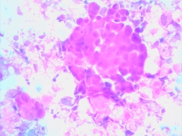
A dying leukemia cell

A dying leukemia cell

The principles underlying the Rational Therapeutics EVA-PCD platform reflect many years of development. Recognizing the importance of cell death measures — apoptotic and non-apoptotic — our laboratory dismissed growth-based assays. The closure of Oncotech, the principal purveyor of proliferation-based assays, illustrates the demise of a failed paradigm in the study and testing of human tumor biology. A second principal of our work is the need to examine all of the operative mechanisms of cell death (autophagic, necrotic, etc.). Laboratories that measure only one mechanism of cell death (e.g. caspase activation as a measure of apoptosis) miss important cell responses that are critical to the accurate prediction of clinical response. The third principle of our work is the maintenance of cells in their native state.

These fundamentals provide the basis of our many successes, but also a constraint. Because we do not propagate, subculture or expand tissues, we can only work with the amounts of tissue provided to us by our surgeons. While some labs propagate small biopsy samples into larger populations by growth to confluence, this introduces irreconcilable artifacts, which diminish the quality of sensitivity profiles. Avoiding this pitfall, however, demands that a tissue sample be large enough (typically 1cm3) to provide an adequate number of cells for study without growth or propagation.

This is the reason our laboratory must request biopsies of adequate size. The old computer dictum of “garbage in, garbage out” is doubly true for small tissue samples. Those that contain too few tumor cells, are contaminated, fibrotic or inadequately processed will not serve the patients who are so desperately in need of therapy selection guidance. As a medical oncologist, I am deeply disappointed by every failed assay and I am more familiar than most with the implications of a patient requiring treatment predicated on little more than intuition or randomization.

We do everything within our power to provide results to our patients. This sometimes requires low yield samples be repeatedly processed. It may also set limitations on the size of the study or, in some circumstances, forces us to report a “no go” (characterized as an assay with insufficient cells or insufficient viability). Of course, it goes without saying that we would never charge a patient for a “no-go” assay beyond a minimal set up fee (if applicable). But, more to the point, we suffer the loss of an opportunity to aid a patient in need.

Cancer patients never undergo therapy without a tissue biopsy. Many have large-volume disease at presentation, so it is virtually always possible to obtain tissue for study if a dedicated team of physicians makes the effort to get it processed and submitted to our laboratory. The time and energy required to conduct an excisional biopsy pales in comparison to the time, energy and lost opportunities associated with months of ineffective, toxic therapy.

Two Women with Metastatic Breast Cancer – Same Age, Same Disease, Two Very Different Functional Profiles

A day in the life of advanced breast cancer. Two different 37-year-old breast cancer patients, both mothers of young children, were seen in consultation on the same day.

The first had been referred by a colleague who was concerned that the patient’s ER positive breast cancer had disseminated to her brain despite aggressive standard chemotherapy. She was to undergo a craniotomy and a portion of fresh tumor would be submitted from the surgery to Rational Therapeutics for EVA-PCD functional profiling.

The second mother had metastatic triple negative breast cancer, which recurred after aggressive standard chemotherapy. She underwent neo-adjuvant treatment (preoperative) but at the time of her surgery, there was no evidence of response to the treatment. By the time we met her, only months into her diagnosis, new areas of metastatic disease were cropping up daily.

Microscope Detail2-lo resThe EVA-PCD assay results on these two “similar” patients were entirely different.

The results of the first patient with the ER positive tumor and brain metastases clearly identified treatments directed toward the PI3K pathway, with or without chemotherapy. We are recommending a combination of Everolimus plus chemotherapy.

The second patient had a completely different profile. Indeed, the degree of drug resistance was quite striking. A three-drug combination was among the most active from almost two dozen drugs tested.  The other option appeared to be a new class of drugs called the cyclin dependent kinase (CDK) inhibitors.

On a functional level, we used targeted drugs to probe for sensitivity to inhibitors of these cancer signal pathways. Unlike genomic profiles that tell you whether the gene is present or absent, we can tell whether the gene is driving the tumor. Functional profiling.

One patient is now under my care and the other will begin treatment under the care of a colleague in Orange County, CA. We will await results of these assay-directed therapies and wish these two young patients every success.

Gastric Cancer: A Call for Patient Selection

Gastric cancer is the fourth most common cancer worldwide with more than 930,000 diagnoses and 800,000 deaths attributed to this disease each year. Although relatively uncommon in the U.S., constituting only 2 percent of new cancers, in countries like Korea it makes up 20 percent of all new malignancies.

Among the causes are Helicobacter pylori infection, diets rich in smoked food, a high intake of nitrates and nitrites and cigarette smoking. A rare but aggressive form of the disease is associated with a gene mutation known as CDH1. The high frequency of metastatic disease at the time of initial diagnosis often precludes surgery, leaving systemic chemotherapy as the principal treatment option.

Annals of Oncology coverA recent report in The Annals of Oncology (No improvement in median survival for patients with metastatic gastric cancer despite increased use of chemotherapy: Bernards N. et al, Annals of Oncology. November, 2013) describes a retrospective analysis by Dutch investigators who examined the use of chemotherapy in patients with inoperable gastric cancer.

In total, 4,797 cases were examined from 1990 to 2011. Over this time, the proportion of patients presenting with metastatic disease increased from 24 percent in 1990 to 44 percent in 2011. At the same time, palliative chemotherapy use increased from 5 percent to 36 percent. Younger patients and those of higher socioeconomic status had the largest increase in chemotherapy use, while older patients, those with linitis plastica and those with multiple metastases had lower chemotherapy use. Despite the significant increase in the use of chemotherapy, the median survival for patients was unchanged at 15 weeks in 1990 and 17 weeks in 2011 (P = 0.1).

Over this period, early treatment regimens like 5-fluorouracil (5FU) and FAM were largely replaced by combinations like Docetaxel/Cisplatin/5-FU (DCF), Cisplatin/Irinotecan, Epirubicin/Oxaliplatin/Capecitabine (EOX) and Carboplatin/Taxol. While response rates and palliative benefits have continued to improve, this has not translated into improved overall survival. This reflects a dilemma that has confronted medical oncologists for decades.

For many years, clinical trialists have held that one cannot assess the benefit of a treatment by comparing responders to non-responders. That is, time to progression and survival must compare all patients on a given treatment arm to those on the control arm. Their rationale was that “one must treat all patients to obtain the benefit seen in some.”  Put differently you cannot “cherry pick” your winners and losers. It was said that this proscription was needed to avoid selection bias. But as any medical or nonmedical person would recognize, people who respond to treatment do better than those who do not. Lacking the ability to identify responders upfront, these trialists have insisted upon a one-size-fits-all approach to the detriment of clinical therapeutics and drug development.

With the dawn of the molecular era we see chinks in the armor of these trial designs as investigators now question why everyone should receive a treatment if only a small percentage will benefit. In gastric cancer, HER2 over-expression, found in 20-25 percent of patients, is now routinely used to identify patients who will respond to trastuzumab. But what of the other 75-80 percent of patients who do not carry HER2 and for whom there are no widely used determinants of clinical response? Do the results of Bernard article suggest that these patients should not receive therapy?

The Bernard article offers an interesting insight into what may be the future of medical oncology. As cancer therapy is increasingly scrutinized, not only for response or palliation but also for overall survival, patients may soon be denied treatments unless the results of the therapy rise to this, the highest level of evidence, for the entire population of treated patients.

Would it not be preferable to use laboratory analyses, like the EVA-PCD®, to select among treatment candidates before subjecting all patients to the risk and expense of toxic chemotherapy? In this regard, the author’s comments are poignant: “Identification of the subgroup of patients which benefit from palliative chemotherapy is of the utmost importance to avoid unnecessary treatment.” As a laboratory investigator engaged in the field of drug selection science (functional profiling), I couldn’t agree more.

Cancer Patients Who Get Better, Get Better

JCO coverA study published in the October 20 Journal of Clinical Oncology (Use of early tumor shrinkage to predict long-term outcome in metastatic colorectal cancer treated with Cetuximab, Piessevaux H. et al, 31:3764-3775,2013) described “early tumor shrinkage” as a predictor of long-term survival in patients with metastatic colorectal cancer. These Belgian and German investigators re-analyzed two large clinical trials in colon cancer, CRYSTAL and OPUS, to evaluate the impact of early tumor shrinkage at eight weeks of therapy. Both studies were in patients with wild type (non-mutated) KRAS colon cancer who received chemotherapy with or without the monoclonal antibody Cetuximab.

They used a cutoff of 20 percent tumor shrinkage at eight weeks to separate “early responders” from “non-responders.” Early responders were found to have a significantly better survival. The accompanying editorial by Jeffrey Oxnard and Lawrence Schwartz (Response phenotype as a predictive biomarker to guide treatment with targeted therapies, J Clin Oncol 31:3739-3741, 2013) examined the implications of this study.

The measurement of tumor response has been a lynchpin of cancer therapeutics for decades. This was later refined under what is known as RECIST (Response Evaluation Criteria In Solid Tumors) criteria. Despite this, there remained controversy regarding the impact of early response on long term survival. The current Piessevaux trial however, is only the most recent addition to a long history of studies that established the correlation between tumor shrinkage and survival. Earlier studies in colorectal, kidney, esophagus and lung cancers have all shown that early response correlates with superior outcomes.

What is gratifying in the accompanying editorial is the discussion of the “response phenotype” as a predictor of survival. Phenotype, defined as “the set of observable characteristics of an individual resulting from the interaction of its genotype with the environment” reflects the totality of human biology not just its informatics (genotype). This renewed appreciation of tumor phenotype in oncology is important for it re-focuses on tumor biology over tumor genetics.

The  ex-vivo analysis of programmed cell death (EVA-PCD) that we utilize, is itself a phenotypic platform that measures actual cellular behavior, not gene profiles, to gauge drug sensitivity. We have previously shown that the measurement of chemotherapy effect on human tumor tissue predicts response, time to progression and survival. The current study used clinical response (early tumor shrinkage) to successfully measure the same.

This analysis of early response by Piessevaux is bringing our most sophisticated investigators back to what they should have known all along.
1. Responding patients do better than non-responding patients.
2. Early measurement of response is predictive of long term outcome.
3. These measurements can and should be done in the laboratory.

Taken together, the current study supports early tumor shrinkage and by inference, ex vivo analyses, as important predictors of patient response and survival.