What Can You Do to Improve Your Odds Against Cancer?

I sometimes joke with my patients that a new diagnosis of cancer rarely provides them enough time to get an MD or PhD. Yet it is that level of preparation that may be required to answer the myriad questions that lie ahead.

Although it’s a joke, it is only partly in jest. Unlike buying a house or a car for which one’s life experiences can prepare you, medicine is opaque, complicated and ever changing. At the bleeding edge of medical complexity sits medical oncology and its dizzying array of genomics, transcriptomics, proteomics, epigenomics and metabolomics. Not only is it difficult for patients to keep up with all the changes, it is increasingly beyond the ken of their doctors who have spent entire careers training in the specialty, many of whom may have an MD and a PhD.

So how can patients improve their odds when the obstacles seem so daunting?

notebook-clipart-red-spiral-notebookMy first recommendation is that you develop a personal diary or record book of the procedures, staging studies, pathologic diagnosis, tumor markers, and physician recommendations. This can be accomplished by requesting that your doctors provide either electronic or physical copies of CT scans, pathology reports, blood tests and other clinically relevant information. While there has been some controversy surrounding their overuse, I am a believer in the simple blood tests used as barometers of your cancer with names like CEA, CA19.9, CA125, CA27.29, and CA 15.3. Although they are not perfect, they are easy to obtain, relatively inexpensive and can be repeated regularly to assess progress with therapy.

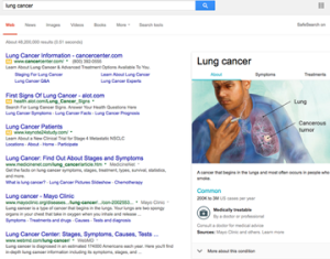
The second thing that I recommend is that you gain a working knowledge of your diagnosis. While there are no lung cancer search resultsperfect sources of information, the internet can provide useful basic information as a starting point. Begin by obtaining from your doctor the most accurate definition of the cancer. If it is breast cancer, is it infiltrating ductal or lobular? Are you ER positive? Is your tumor HER-2 positive? If it is stomach cancer, is it intestinal type or diffuse, etc? This will facilitate your searches, as well as your future conversations with consultants.
Once you know what you’ve got, the next thing you will need to know is where it is. That is what is known as your stage. The older classification used Roman Numerals I-IV with local disease (early) as stage I and metastatic (disseminated) as stage IV. The more modern system is known as TNM, where T stands for tumor size (1-4), N stands for lymph involvement(1-3), and M stands for metastatic involvement (0 or 1). Most contemporary pathology reports include TNM staging. With the diagnosis and stage established, you now know what you have and where it is.

This is where it gets interesting. Now, what do you do about it?

It is at this point that therapeutic choices must be made. Most physicians will rely upon standard established guidelines. Among the most widely used guidelines are those published by the National Comprehensive Cancer Network known as NCCN. While these guidelines can be useful, they can also be stultifying, limiting patients to what might be considered the lowest common denominator of care. While they may be better than haphazard treatment selection, they may very much miss the mark for your unique needs.

Here the process degenerates into a plethora of confusing choices.

Should you have genomic profiling? If so, should it be based on a tissue biopsy, circulating cell free tumor analysis, or even the newer urine tests that measure the presence or absence of abnormal genes? All of these technologies have merit and over the coming years the best ones will shake out. Despite these tests being widely touted (and profitable for the purveyors), none of these test have been put to formal trials that establish their capacity to influence survival. This is interesting because many of these tests have obtained insurance and Medicare coverage without even remotely rising to this standard. Nonetheless, these tests can be used for specific diseases like lung and leukemia where actionable targets are known to exist. Beyond that, caveat emptor (buyer beware).

One of the problems with genomic profiles is that they do very good job of telling you what the problem may be, but a very bad job at telling you the solution. It is a rare genomic mutation that comes with a drug to treat it. Most of the findings wind up asking more questions rather than providing more answers.

With the diagnosis established, the stage known and in certain circumstances molecular profiles complete, it is time for you to choose treatments and the centers that will provide them. Many seek the care of academic centers. These centers may offer clinical trials as a first line therapy for those who meet criteria.
NCI Clinical TrialsIt should be remembered that clinical trials are conducted in three principal formats. Phase I trials examine brand new drugs. These trials determine the safety of the drugs at different dose schedules. Phase II trials take the established safe doses and develop experience in each type of disease, e.g. lung versus colon versus breast. Phase III trials then compare the new drugs with existing treatments to see if there is any real improvement.

It is critical to recognize the functions of these different types of trials. Phase I studies classically have no therapeutic intent (your benefit is secondary to their measurement of your ability to tolerate the drug).

Phase II trials seek evidence of clinical activity by disease, but your specific disease may not be right for that drug.

Finally, Phase III allows a comparison of standard treatment to the new one. Many of these drugs do not make the grade and fall off the development wagon. In addition, you must be willing to be randomly assigned.

It is here that my approach diverges from those outlined. I have long maintained that each patient is unique and that their cancers must be treated individually. Recognizing that no genomic, proteomic or transcriptomic platform can answer the very complex questions of therapeutic response, we at Rational Therapeutics have developed functional analyses through the use of the EVA-PCA assay, which studies each patient’s tumor by exposing it to the drugs of interest. The most active, least toxic combinations are then recommended. In a report at the American Society of Clinical Oncology meeting of 2013, we showed a 2.02 higher response rate (P < 0.001) and a 1.44 improvement in one year survival (P < 0.02) for patients who received assay-guided therapy. This established the predicative validity of the functional approach.

It is important for patients to realize that cancer is an unbalanced system, not just an abnormal cell. Cancer as a disease goes beyond the cell or even the tumor to affect the body itself. Alterations in immunity, metabolism and physiology contribute to the good or bad outcomes of every patient. Patients should seek to normalize their lifestyle, improve their diets, maintain an active exercise program, reduce their weight to lean body weight, and may in some circumstances consider nutritional supplements and/or appropriately selected natural products that may augment their wellbeing.

The human body is a complicated machine and each part resonates with every other part. A good diet, a good night’s sleep and avoidance of an unhealthy lifestyle, as much as they may sound like your mother’s advice, is indeed very good advice.

Every cancer patient has the right to get better. As a patient, you should take charge of your cancer and make smart decisions. Afterall, no one is more interested in saving your life than you.

Investigators in Boston Re-Invent the Wheel

A report published in Cell from Dana-Farber Cancer Institute describes a technique to measure drug cov150hinduced cell death in cell lines and human cancer cells. The method “Dynamic BH3 profiling” uses an oligopeptidic BIM to gauge the degree to which cancer cells are “primed” to die following exposure to drugs and signal transduction inhibitors. The results are provocative and suggest that in cell lines and some human primary tissues, the method may select for sensitivity and resistance.

We applaud these investigators’ recognition of the importance of phenotypic measures in drug discovery and drug selection and agree with the points that they raise regarding the superiority of functional platforms over static (omic) measures performed on paraffin fixed tissues. It is heartening that scientists from so august an institution as Dana-Farber should come to the same understanding of human cancer biology that many dedicated researchers had pioneered over the preceding five decades.

Several points bear consideration. The first, as these investigators so correctly point out: “DBP should only be predictive if the mitochondrial apoptosis pathway is being engaged.” This underscores the limitation of this methodology in that it only measures one form of programmed cell death – apoptosis. It well known that apoptosis is but one of many pathways of programmed cell death, which include necroptosis, autophagy and others.

While leukemias are highly apoptosis driven, the same cannot so easily be said of many solid tumors like colon, pancreas and lung. That is, apoptosis may be a great predictor of response except when it is not. The limited results with ovarian cancers (also apoptosis driven) are far from definitive and may better reflect unique features of epithelial ovarian cancers among solid tumors than the broad generalizability of the technique.

A second point is that these “single cell suspensions” do not recreate the microenvironment of human tumors replete with stroma, vasculature, effector immune cells and cytokines. As Rakesh Jain, a member of the same faculty, and others have so eloquently shown, cancer is not a cell but a system. Gauging the system by only one component may grossly underestimate the systems’ complexity, bringing to mind the allegory of elephant and the blind man. Continuing this line of reasoning, how might these investigators apply their technique to newer classes of drugs that influence vasculature, fibroblasts or stroma as their principal modes of action? It is now recognized that micro environmental factors may contribute greatly to cell survival in many solid tumors. Assay systems must be capable of capturing human tumors in their “native state” to accurately measure these complex contributions.

Thirdly, the ROC analyses consistently show that this 16-hour endpoint highly correlates with 72- and 96-hour measures of cell death. The authors state, “that there is a significant correlation between ∆% priming and ∆% cell death” and return to this finding repeatedly. Given that existing short term (72 – 96 hour) assays that measure global drug induced cell death (apoptotic and non-apoptotic) in human tumor primary cultures have already established high degrees of predictive validity with an ROC of 0.89, a 2.04 fold higher objective response rate (p =0.0015) and a 1.44 fold higher one-year survival (p = 0.02) are we to assume that the key contribution of this technique is 56 hour time advantage? If so, is this of any clinical relevance? The report further notes that 7/24 (29%) of ovarian cancer and 5/30 (16%) CML samples could not be evaluated, rendering the efficiency of this platform demonstrably lower than that of many existing techniques that provide actionable results in over 90% of samples.

Most concerning however, is the authors’ lack of recognition of the seminal influence of previous investigators in this arena. One is left with the impression that this entire field of investigation began in 2008. It may be instructive for these researchers to read the first paper of this type in the literature published in in the JNCI in 1954 by Black and Spear. They might also benefit by examining the contributions of dedicated scientists like Larry Weisenthal, Andrew Bosanquet and Ian Cree, all of whom published similar studies with similar predictive validities many years earlier.

If this paper serves to finally alert the academic community of the importance of human tumor primary culture analyses for drug discovery and individual patient drug selection then it will have served an important purpose for a field that has been grossly underappreciated and underutilized for decades. Mankind’s earliest use of the wheel dates to Mesopotamia in 3500 BC. No one today would argue with the utility of this tool. Claiming to have invented it anew however is something quite different.

A New Use for One of the Oldest “New” Drugs

With the profusion of new targeted agents entering the clinical arena, a report from the American Society of Hematology bears consideration.

The trial known as the SORAML trial enrolled 276 patients with newly diagnosed acute myelogenous leukemia. The patients were between the ages of 18 and 60. All patients received a standard chemotherapy regimen. The patients were then randomized to receive Sorafenib or placebo. Patients on the Sorafenib arm then remained on a maintenance therapy for twelve months.

While the achievement of complete remission was almost identical between the two arms at 59% and 60%, the event free survival demonstrably favored the Sorafenib group at 20.5 months versus 9.2 months. At three years of follow-up 40% of the Sorafenib group were well with only 22% of the placebo group still in remission. This corresponds to a three-year relapse free survival of 38% for placebo and 56% for Sorafenib (P=0.017).

The results are of interest on several levels.
1.    Sorafenib a multitargeted tyrosine kinase inhibitor was approved in December 2005 for the treatment of renal cell carcinoma. This makes Sorafenib one of the first targeted agents to achieve FDA approval.

2.     Sorafenib has many modes of action and it is not entirely clear which of its functions were responsible for the superior survival in this AML study.

3.    Sorafenib’s approval reflects a rather convoluted and interesting history. When first developed the drug was designed to target the oncogene B-Raf. As a result the drug was introduced into early clinical trials for the treatment of advanced melanoma, a disease known to be associated with B-Raf mutation. As the drug proved ineffective, it appeared unlikely to gain FDA approval. That is, until it showed cross reactivity with VEGF pathway associated with tumor cell vascularity. A successful trial published in the New England Journal of Medicine then led to the approval.

Now, nine years later this old new drug has gained new life. This time in acute myelogenous leukemia.

The term “dirty drug” refers to agents that target many kinases at the same time. Sorafenib is an example of a “dirty drug.” However it is Sorafenib’s “dirty drug” quality that led first to its approval and most likely now leads to its application in AML. This reflects the fact that Sorafenib may be inhibiting B-Raf signaling associated with the common mutation in Ras upstream of B-Raf or it may reflect Flt3 a secondary activity associated with Sorafenib.

Indeed B-Raf and Flt3 may not be upregulated in every patient, but could serve a function of permissive activity granting an additional survival signal to the AML cells as they go through induction therapy. These subtleties of drug effect may escape genomic analysis as the true “target” may not be mutated, upregulated or amplified. No doubt the investigators in this study will conduct gene sequencing to determine whether there is a driver mutation associated with the advantage reported in this clinical study. What will be intriguing is to determine whether that advantage is an abnormal gene functioning within these cancerous cells or possibly a normal gene functioning abnormally in these cancer cells. More to come.

Genomic Profiling for Lung Cancer: the Good, the Bad and the Ugly

Genomic profiling has gained popularity in medical oncology. Using NextGen platforms, protein coding regions of human tumors known as exomes can be examined for mutations, amplifications, deletions, splice variants and SNPs. In select tumors the results can be extremely helpful. Among the best examples are adenocarcinomas of the lung where EGFr, ALK and ROS-1 mutations, deletions and/or re-arrangements identified by DNA analysis can guide the selection of “targeted agents” like Erlotinib and Crizotinib.

An article published in May 2014 issue of JAMA reported results using probes for 10 “oncogenic driver” mutations in lung cancer patients. They screened for at least one gene in 1,007 patients and all 10 genes in 733. The most common was k-ras at 25%, followed by EGFR in 17% and ALK in 8%. The incidence then fell off with other EGFr mutations in 4%, B-raf mutations in 2%, with the remaining mutations each found in less than 1%.

Median survival at 3.5 vs 2.4 years was improved for patients who received treatments guided by the findings (Kris MG et al, Using multiplex assays of oncogenic drivers in lung cancers to select targeted drugs. JAMA, May 2014). Do these results indicate that genomic analyses should be used for treatment selection in all patients? Yes and no.

Noteworthy is the fact that 28% of the patients had driver mutations in one of three genes, EGFr, HER2 or ALK. All three of these mutations have commercially available chemotherapeutic agents in the form of Erlotinib, Afatinib and Crizotinib. Response rates of 50% or higher, with many patients enjoying durable benefits have been observed. Furthermore, patients with EGFr mutations are often younger, female and non-smokers whose tumors often respond better to both targeted and non-targeted therapies. These factors would explain in part the good survival numbers reported in the JAMA article. Today, a large number of commercial laboratories offer these tests as part of standard panels. And, like k-ras mutations in colon cancer or BCR-abl in CML (the target of Gleevec), the arguments in favor of the use of these analyses is strong.

Non-small cell lung cancer

Non-small cell lung cancer

But what of the NSCLC patients for whom no clear identifiable driver can be found? What of the 25% with k-ras mutations for whom no drug exists? What of those with complex mutational findings? And finally what of those patients whose tumors are driven by normal genes functioning abnormally? In these patients no mutations exists at all. How best do we manage these patients?

I was reminded of this question as I reviewed a genomic analysis reported to one of my colleagues. He had submitted a tissue block to an east coast commercial lab when one of his lung cancer patients relapsed. The results revealed mutations in EGFr L858R & T790M, ERBB4, HGF, JAK2, PTEN, STK11, CCNE1, CDKN2A/B, MYC, MLL2 W2006, NFKB1A, and NKX2-1. With a tumor literally bristling with potential targets, what is a clinician to do? How do we take over a dozen genetically identified targets and turn them into effective treatment strategies? In this instance, too much information can be every bit as paralyzing as too little.

Our preferred approach is to examine the small molecule inhibitors that target each of the identified aberrancies in our laboratory platform. We prefer to drill down to the next level of certainty e.g. cellular function. After all, the presence of a target does not a response make.

In this patient I would conduct a biopsy. This would enable us to examine the drugs and combinations that are active against the targets. A “hit” by the EVA-PCD assay would then isolate the “drivers” from the “passengers” and enable the clinician to intelligently select effective treatments. Combining genomic analyses with functional profiling (phenotypic analyses) provides the opportunity to turn speculative observations into actionable events.

This is the essence of Rational Therapeutics.

The Frustrating Reality – When a Tumor Sample isn’t Sufficient for Testing

A dying leukemia cell

A dying leukemia cell

The principles underlying the Rational Therapeutics EVA-PCD platform reflect many years of development. Recognizing the importance of cell death measures — apoptotic and non-apoptotic — our laboratory dismissed growth-based assays. The closure of Oncotech, the principal purveyor of proliferation-based assays, illustrates the demise of a failed paradigm in the study and testing of human tumor biology. A second principal of our work is the need to examine all of the operative mechanisms of cell death (autophagic, necrotic, etc.). Laboratories that measure only one mechanism of cell death (e.g. caspase activation as a measure of apoptosis) miss important cell responses that are critical to the accurate prediction of clinical response. The third principle of our work is the maintenance of cells in their native state.

These fundamentals provide the basis of our many successes, but also a constraint. Because we do not propagate, subculture or expand tissues, we can only work with the amounts of tissue provided to us by our surgeons. While some labs propagate small biopsy samples into larger populations by growth to confluence, this introduces irreconcilable artifacts, which diminish the quality of sensitivity profiles. Avoiding this pitfall, however, demands that a tissue sample be large enough (typically 1cm3) to provide an adequate number of cells for study without growth or propagation.

This is the reason our laboratory must request biopsies of adequate size. The old computer dictum of “garbage in, garbage out” is doubly true for small tissue samples. Those that contain too few tumor cells, are contaminated, fibrotic or inadequately processed will not serve the patients who are so desperately in need of therapy selection guidance. As a medical oncologist, I am deeply disappointed by every failed assay and I am more familiar than most with the implications of a patient requiring treatment predicated on little more than intuition or randomization.

We do everything within our power to provide results to our patients. This sometimes requires low yield samples be repeatedly processed. It may also set limitations on the size of the study or, in some circumstances, forces us to report a “no go” (characterized as an assay with insufficient cells or insufficient viability). Of course, it goes without saying that we would never charge a patient for a “no-go” assay beyond a minimal set up fee (if applicable). But, more to the point, we suffer the loss of an opportunity to aid a patient in need.

Cancer patients never undergo therapy without a tissue biopsy. Many have large-volume disease at presentation, so it is virtually always possible to obtain tissue for study if a dedicated team of physicians makes the effort to get it processed and submitted to our laboratory. The time and energy required to conduct an excisional biopsy pales in comparison to the time, energy and lost opportunities associated with months of ineffective, toxic therapy.

The False Economy of Genomic Analyses

While Dr. Nagourney is enjoying Spring Break with his family, here is a very topical subject in light of the constant press given to genetic testing.

Dr. Robert A. Nagourney - Rational Therapeutics - Blog

We are witness to a revolution in cancer therapeutics. Targeted therapies, named for their capacity to target specific tumor related features, are being developed and marketed at a rapid pace. Yet with an objective response rate of 10 percent (Von Hoff et al JCO, Nov 2011) reported for a gene array/IHC platform that attempted to select drugs for individual patients we have a long way to go before these tests will have meaningful clinical applications.

So, let’s examine the more established, accurate and validated methodologies currently in use for patients with advanced non-small cell lung cancer. I speak of patients with EGFR mutations for which erlotinib (Tarceva®) is an approved therapy and those with ALK gene rearrangements for which the drug crizotinib (Xalkori®) has recently been approved.

The incidence of ALK gene rearrangement within patients with non-small cell lung cancer is in the range of 2–4 percent, while EGFR mutations…

View original post 396 more words

Our Blog’s 2013 Year in Review

WordPress.com came up with this nifty report of our blog’s progress for 2013. We thought we’d share it with you. Do you have anything to add? Tell us what you would like to see during the coming new year.

Wishing you good health and much happiness in 2014.

Here’s an excerpt:

The concert hall at the Sydney Opera House holds 2,700 people. This blog was viewed about 22,000 times in 2013. If it were a concert at Sydney Opera House, it would take about 8 sold-out performances for that many people to see it.

Click here to see the complete report.

Chemosensitivity Testing Captures Attention of “Nature Biotechnology”

Nature Biotech largecoverAn interesting editorial appeared in the February 2013 issue of Nature Biotechnology titled “Dishing out cancer treatment.” The lead line reads, “Despite their limitations, in-vitro assays are a simple means for assessing the drug sensitivity of a patient’s cancer . . . we think assays deserve a second look.”

The author describes the unequivocal appeal of laboratory analyses that are capable of selecting drugs and combinations for individual patients. At a time when 100’s of new drugs are in development, drug discovery platforms that can mimic human tumor response in the laboratory are becoming increasingly attractive to patients and the pharmaceutical industry. While the author, rooted in contemporary molecular biology, examines the field through the lens of genomic, transcriptomic, proteomic and metabolomic profiling, he recognizes that these analyte-based approaches cannot capture the tumor in its microenvironment, yet we now recognize that these micro-environmental influences are critical to accurate response prediction.

As one reads this piece, it is instructive to remember that no other platform can examine the dynamic interaction between cells and their microenvironment. No other platform can examine drug synergy. And no other platform can examine drug sequence.

It is these complexities however, that will guide the next generation of drug tests and ultimately the process of drug discovery. Even the most ardent adherents to genomic profiling must ultimately recognize that genotype does not equal phenotype. Yet, it is the tumor phenotype that we must study.

I am gratified that the editors of so august a journal as Nature Biotechnology have taken the time to reexamine this important field. Perhaps, if our most scientific colleagues are beginning to recognize the importance of functional analyses, it may be only a matter of time before the clinical oncology community follows suit.

The editor’s final line is poignant, “After years spent on the sidelines, perhaps in-vitro screening methods deserve another look.” We couldn’t agree more.

What Goes Around Comes Around

On a recent morning, I found myself listening to an analysis of the public defender program in America. It seems that defendants who are provided with publically funded lawyers are almost uniformly advised to submit pleas. Regardless of the strength of their cases or their apparent innocence, plea bargains have become the preferred legal defense.

As I listened, the reason became increasingly apparent. It seems that in many states, legal fees paid to public defenders are bundled into a single, one-time payment. Lawyers are provided the same amount of money whether the client is convicted or found innocent. If the lawyers choose to vigorously defend their clients and prove their innocence, they receive precisely the same amount of money that they would receive if the accused pleaded guilty on the spot. It has not taken long for the legal community to figure out that plea bargains are good for business.

As a physician, I found this analysis of more than passing interest. After all, the movement toward managed care and the DRG (diagnosis-related groups) system make lump sum payments for medical care the norm. Using arbitrary metrics like per member/per month, physicians receive the same amount of the money whether they save your life or not. The defenders of this approach explain that it was designed to limit the profit motive and curtail physician avarice. What it has succeeded in doing however, is to incentivize physicians to give up. If there is no return on investment, then there will be no investment of time, energy, skill or ability. If physicians receive no more payment, accolades, or recognition for good outcomes than for bad ones, how long can we realistically expect good outcomes to continue?

The district attorneys, who were interviewed for the piece on public defenders, pointed out, in their defense, that this has succeeded in “clearing the dockets” of backlogged court cases. However, if we were to extend that line of reasoning into the realm of medical care delivery, we would need to confront a number of distinctly unappealing ramifications.

The corporatization of medicine, expanding regulation, creation and promotion of the HMO model, and recent legislation are all the products of legal minds. While NCCN cancer therapy guidelines are increasingly employed, not as guidelines, but instead as dictates to limit the ability of well-trained, sophisticated oncologists to practice their art; a recent article in the New England Journal of Medicine on bending the healthcare curve had the “underutilization” of hospice care as a principal focus. It is interesting to observe how lawyers, faced with “lump sum” payments, are responding. Is the reported deterioration of legal representation, especially for the under privileged in America, the jurisprudence counterpart to policies already underway in the medical system? Will the same disincentives in place for physicians now come home to roost for lawyers?

Cancer Gets Personal

Early in the morning of Nov 21, I suffered the loss of my father. However prepared one might be for this eventuality, there is nothing that can really prepare you.

At 95 years of age, he had lived longer than many. I had cared for my father as a patient since 1974, when he was first diagnosed with inoperable prostate cancer. I remember the day I received notification of the diagnosis. I felt a sense of deep sorrow that my father at 74 would soon die of high-grade locally advanced prostate carcinoma. As a member of the generation that forgot to have children, I was saddened that my father would not live to see grandchildren.

I remember traveling to Connecticut for his initial evaluation and then scouring the literature for the best possible options. Fortunately, despite the aggressiveness of the disease it had not metastasized.

I arranged for my father to travel to California where I then oversaw his care in collaboration with Dr. A.M. Nisar Syed in radiation oncology. There is a well-known dictum in medicine that only doctors and their families suffer unexpected complications. In my father’s case it certainly rang true. First, the radiation implants did not penetrate the tumor and needed to be removed and replaced. As a result of this double procedure, he then developed bleeding that required emergency hospitalization several days later.

Despite these hiccups, the combination of implant and external beam radiation provided excellent control. With a full recovery my father returned to his normal activities.

As so often happens in medicine a personal experience provides a focused interest. I delved into the prostate cancer literature and became increasingly interested in the biology of this disease. One area of particular interest was the role of hormonal therapy. When? How much? How long?

When my father’s PSA began to rise the second year, I had a unique opportunity to examine these questions at a very personal level. Would the early institution of androgen blockade induce the hormone refractory state? Was there a “trigger” value of the PSA that dictated the institution of the therapy? I remember discussing these questions with a prostate cancer expert and chairman of the ECOG committee, Dr. Basil Kasimis, whom I had had the pleasure of working with several years earlier. I agonized over starting hormonal therapy as my father’s PSA rose from 4 to 10, to 25, to 54, and up to 150. Despite these frightening PSA values, there was no evidence of metastatic disease on serial bone scans, which I performed religiously every six to 12 months.

Almost a decade passed but there was still no metastatic disease. And then my father developed severe coronary artery disease in his early 80s. Coronary artery bypass graft was the only option. To avoid the possibility of seeding the sternal wound, I bit the bullet and treated him with hormonal suppression – immediately driving the PSA to nearly 0.

With his coronary artery bypass surgery a success, he came off hormonal therapy and I let his PSA drift upward again.

As he had returned to Connecticut, his urologist became increasingly concerned by the rising PSA, and, without my knowledge, decided to rechallenge him with hormonal ablation. While I understood the motivation for this intervention, I didn’t agree and took him off all hormones for a prolonged period of time. Over the subsequent years, I would intervene occasionally to shepherd my father through pneumonia, a broken hip, a bleeding ulcer, and a variety of other maladies so common in patients who transition from their 80s to their 90s. On several occasions, we gave brief courses of hormonal ablation to suppress the PSA, when the steepness of the rise gave concern. Twenty-one years after his diagnosis my father died of natural causes, with no evidence of metastatic prostate cancer.

The experience was instructive on many levels. First, I realized how important it is to treat all patients as if they are a member of your own family. Second, it takes a lot of guts to step outside the normal guidelines and to do what you believe to be best. Third, I realize that in medical oncology it is the most “aggressive” physician who has the courage not to treat.

So often in this field doctors institute treatment, not because it is needed, nor because it will work, but because by doing so they have “done their job,” the rest is no longer their responsibility.

But “doing your job” as a physician, particularly in medical oncology may demand that you step outside of the NCCN guidelines, however uncomfortable it may make you, to do the right thing. Virtually every urologist or oncologist in America would have treated my father for his rising PSA 20 years ago. While I cannot say with certainty, I feel fairly confident that he lived the past 21 years in part because I didn’t treat him. Every patient needs an advocate. I feel a sense of personal satisfaction that I was there to be my father’s. He lived a long and productive life, I hope and believe that I helped him to do so.

Every experience, even traumatic ones, can have a silver lining. My father’s diagnosis lead me to develop a combined modality approach for locally advanced prostate cancer that has provided among the best biochemical relapse-free survival rates ever observed in this disease. Had I known then what I know today, I would have certainly treated my father with this approach.

Secondly, my interest in prostate cancer lead me to examine the lifestyle, nutritional, and micro-nutritional aspects of this disease – knowledge that I apply to this day. This lead to my analysis of an herbal remedy for prostate cancer that unfortunately uncovered the adulteration of an herbal mixture as we reported. (Herbal Composition PC-SPES for Management of Prostate Cancer: Identification of Active Principles: Journal of National Center Institute, Vol. 94, No. 17, September 4, 2002.) Despite our disappointment at the discovery, it lead me to reexamine the use of estrogenic substances as therapies in this disease, insights that have provided benefit to many of my patients ever since.

In retrospect, it may have been my father’s natural inquisitiveness (that he imparted to me) that leads to my pursuit of these lines of investigation. And for that I will always be grateful.

To read more about Alphonse Nagourney, click here.