What is Personalized Cancer Therapy?

Personalized therapy is the right treatment, at the right dose for the right patient. Like the weather, however, it seems that everyone’s talking about it, but no one is doing anything about it.

In its simplest form personalized care is treatment that is designed to meet an individual’s unique biological features. Like a key in a lock, the right drug or combination opens the door to a good outcome.

Lock & Keys 1When over the years I lectured on the development of the cisplatin/gemcitabine doublet, my two boys were quite young. I would show a slide depicting a doorknob with a key in the keyhole. I likened our lab’s capacity to identify sensitivity to the cisplatin/gemcitabine combination as “unlocking” an individual’s response.

At the time my wife and I would leave the key in the inside of the front door enabling us to unlock it when going out. We reasoned at the time that our 2-year-old would not be strong enough, nor tall enough to turn the key and let himself outside. We reasoned wrong, for one day our son Alex reached up, turned the key and opened the door right in front of us. Lesson learned: Given the right key, anyone can open a door.

I continued my analogy by saying that even Arnold Schwarzenegger would be unable to open a door given the wrong key, but might, if he continued trying, snap it off in the lock.

The right key is the right treatment, effortlessly unlocking a good response, while the wrong key is the wrong treatment more often than not too much, too late, akin to a solid tumor bone marrow transplant.

In recent years, personalized care has come to be considered synonymous with genomic profiling. While we applaud breakthroughs in human genomics today, there is no molecular platform that can match patients to treatments. The objective response rate of just 10 percent, almost all in breast and ovarian cancer patients in one study (Von Hoff J Clin Oncol 2010 Nov 20:28(33): 4877-83), suggests that cancer biology is demonstrably more complex than an enumeration of its constituent DNA base pairs. The unilateral focus on this area of investigation over others might be described as “the triumph of hope over experience” (James Boswell, Life of Samuel Johnson, 1791).

But hope springs eternal and with it the very real possibility of improving our patients outcomes. By accepting, even embracing, the complexity of human tumor biology we are at the crossroads of a new future in cancer medicine.

William Withering (1741-1799) the English physician and botanist credited with discovering digitalis as the therapy for dropsy, e.g. congestive heart failure (An Account of the Foxglove and some of its Medical Uses, Withering W. 1785), had absolutely no idea what a membrane ATPase was, when he made his remarkable discovery. It didn’t matter. Cardiac glycosides provided lifesaving relief to those who suffered from this malady for fully two centuries before Danish scientist, Jens Christian Skou, identified these membrane bound enzymes, for which he was awarded a Nobel Prize in 1997.

Similarly, penicillin, aspirin, and morphine were in all use for decades, centuries, even millenia before their actual modes of action were unraveled. Medical doctors must use any and all resources at their disposal to meet the needs of their patients. They do not need to know “how” something works so much as they (and their patients) need to know “that” it works.

The guiding principle of personalized medicine is to match patients to therapies. Nowhere in this directive is there a prescription of the specific platform to be used. Where genomic signatures provide useful insights for drug selection, as they do in APL (ATRA, Arsenic trioxide); NSCLC (EGFr, ROS1, ALK); CML (Imatinib, Dasatanib) then they should be used.

However, in those disease where we haven’t the luxury of known targets or established pathways, i.e. most human malignancies, then more global assessments of human tumor biology should, indeed must, be used if we are to meet the needs of our patients. Primary culture analyses like the EVA-PCD® provide a window onto human tumor biology. They are vehicles for therapy improvement and conduits for drug discovery. Scientists and clinicians alike need to apply any and all available methodologies to advance their art. The dawn of personalized medicine will indeed be bright if we use all the arrows in our quiver to advance clinical therapeutics and basic research.

Reposted from May 2012
Cancer Research Moves Forward by Fits and Starts

AACR logoI recently returned from the American Association for Cancer Research (AACR) meeting held in Philadelphia. AACR is attended by basic researchers focused on the molecular basis of oncology. Many of the concepts reported will percolate to the clinical literature over the coming years.

There were many themes including the revolution in immunologic therapy that took center stage, as James Allison, PhD, received the Pezcoller Prize for his groundbreaking work in targeting immune checkpoints. The Princess Takamatsu Award given to Dr. Lewis Cantley, recognized his seminal contribution to our understanding of signal transduction at the level of PI3K. A series of very informative lectures were provided on “liquid biopsies” that examine blood, serum and other bodily fluids to characterize the process of carcinogenesis. These technologies have the potential to revolutionize the diagnosis and monitoring of cancers.

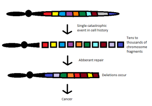
The first symposium I attended described the phenomenon of chromothripsis. This represents a catastrophic cellular trauma that results in the simultaneous fragmentation of chromosomal regions, allowing for rejoining of disparate chromosome components, often leading to malignancy and other diseases. I find the concept intriguing, as it reflects the intersection of oncology with evolutionary developmental biology, reminiscent of the outstanding work of Stephen Jay Gould. His theory of punctuated equilibrium, from 1972, challenged many long held beliefs in the study of evolution.

Since the time of Charles Darwin, we believed that evolution was slow and continual.  New attributes were selected under environmental pressure and the population carried those characteristics forward toward higher complexity. Gould and his associate, Niles Eldredge, stated that evolution was anything but gradual. Indeed, according to their hypothesis, evolution occurred as a state of relative stability, followed by brief episodes of disruption. This came to mind as I contemplated the implications of chromothripsis.

Licensed under CC BY-SA 3.0 via Wikimedia Commons

Licensed under CC BY-SA 3.0 via Wikimedia Commons

According to the new thinking (chromothripsis and its related fields), cancer may arise as a single cell forced to recover from what would otherwise be catastrophic injury. The reconfiguring of genetic elements scrambled together to avoid apoptosis (programmed cell death) provides an entirely new biology that can progress to full-blown malignancy.

By this reasoning, each patient’s cancer is unique. The results of damage control whereby chromosomal material is rejoined haphazardly would be largely unpredictable. These cancers would have a fingerprint all their own, depending on which chromosome was disrupted.

As high throughput technologies and next generation sequences continue to unravel the complexity of human cancer, we seem to be more and more like those who practice stone rubbing to create facsimiles of reality from the “surface” of our genetic information. Like stone rubbing, practitioners do not create the images, but simply borrow from them.

With each symposium, we learn that cancer biology does not come to be, but is. Grasping the complexity of cancer requires the next level of depth. That level of depth is slowly being recognized by investigators from Harvard University to Vanderbilt as the measurement of humor tumor phenotypes.

Cancer is phenotypic and human biology is phenotypic. Laboratory analyses that allow us to measure, grasp, and manipulate phenotypes are those that will provide the best outcomes for patients. Laboratory analyses like the EVA-PCD.

Breakthroughs In Cancer?

Coco Chanel, the icon of 20th century fashion once said, “Only those with no memory insist on their originality.” I am reminded of this quote as I review recent discoveries in cancer, among them, the recognition that cancer represents a dysregulation of cellular metabolism.

The field of metabolomics (the systematic study of cellular energy production), explored by investigators over the last decade is little more than the rediscovery of enzymology (a branch of biochemistry that deals with the properties, activity, and significance of enzymes), biochemistry (the science dealing with the chemistry of living matter) and stoichiometry (the part of chemistry that studies amounts of substances that are involved in reactions), pioneered by investigators like Albert Lehninger, Hans Krebs, Otto Warburg, and Albert Szent-Gyorgyi. These innovators used crude tools to explore the basis of human metabolism as they crafted an understanding of bioenergetics (the study of the transformation of energy in living organisms) and oxidative phosphorylation (processes occurring in the cell’s mitochondrion that produce energy through the synthesis of ATP (energy carrier of the body).

More recently, scientists wedded to genomics have slowly come to recognize the limitations of their approach and have returned to the field of phenotypic (the observable physical or biochemical characteristics of an organism analysis.

While newcomers to the field claim to be the first to recognize the role of cellular biology in tumor biology, a cadre of dedicated investigators had already charted these waters decades earlier. Beginning with the earliest studies by Siminovitch, McCulloch and Till, subsequent investigations by Sydney Salmon and Anne Hamburger, developed the earliest iteration of cellular studies for the examination of cancer biology in primary culture.

Ovarian Cancer

Ovarian Cancer

The work of Black and Spear, published in the 1950s similarly explored the study of human cellular behavior for the study of cancer research. While Larry Weisenthal, Andrew Bosanquet and others established useful predictive methodologies to study cellular phenotype, their seminal contributions have gone largely unrecognized.

Today, start-up companies are examining cellular biology to predict cancer outcomes, each claiming to be the first to recognize the importance of cell death events in primary culture. The most recent and widely touted in the literature is the use of mouse avatars. Implanting biopsied explants of tissue from patients into nude mice, they grow the cancers to desired size and then inject the drugs of interest to show tumor shrinkage. To the discerning eye however, it obvious that this represents little more than an expensive, inefficient, and extremely slow way to achieve that, which can be done more easily, inexpensively, and quickly in a tissue culture environment.

When I read the promotional material of some of the new entrants to this field, I am reminded of another quote, that of Marie Antoinette, who said, “There is nothing new except what has been forgotten.”

The Case for the Metabolic Basis of Cancer Gains Traction

Researchers from the Huntsman Cancer Institute at the University of Utah reported an interesting finding with far-reaching implications.

In their study of the rare tumor known as alveolar soft part sarcoma (ASPS), they examined the well-established chromosomal translocation that occurs between chromosomes 17 and X. This results in the 250px-Protein_ASPSCR1_PDB_2al3production of a fusion protein dubbed ASPSCR1-TFE3. Like other fusion proteins described in malignancies such as lymphoma, acute pro-myelocytic leukemia and chronic myelogenous leukemia, a novel function occurs when two disparate genomic elements are spliced together.

In this instance, the ASPSCR1-TFE3 gene product functions as a lactate transporter. Strikingly, every mouse in which the gene was up regulated developed a tumor. The locations of tumor, in the skull and near the eye, both represented areas of high lactate concentration. In humans, this tumor occurs in skeletal muscle, also associated with high lactate production.

Since 1930, when Otto Warburg first described increased glycolysis (preferential use of sugars) in tumor cells, investigators have pondered the implication of inefficient glucose metabolism in the face of adequate oxygenation.

Human metabolism relies upon mitochondrial function to efficiently liberate the maximum amount of energy in the form of ATP from each glucose molecule. Glycolysis occurring in the cell cytoplasm is highly inefficient and produces only 1/18 of the amount of ATP that a full molecule of glucose can produce through mitochondrial oxidative phosphorylation. Recent molecular biological studies have established that the preferential use of glycolysis may represent the cells need to direct glucose away from energy production and toward the creation of essential structures like amino acids, lipids, and nucleic acids. With the rapid turnover of glucose, cells produce an overwhelming amount of lactate, which is then transported out of the cell. At least this has been the working hypothesis over many years.

More recently, investigators have begun to examine how lactate metabolism may represent the interplay between stromal fibroblast cells and tumor cells. Indeed, many tumor cells are now known to increase lactate uptake reflecting increased lactate production by fibroblasts that have been commandeered in the tumor microenvironment.

Lactate uptake is under the control of a family of transporters known as monocarboxylate transporters, of which nine have been described. These are expressed differently in various tissues, have different affinities for lactate and transport in one direction or another. These processes appear to be under the control of the major regulator of oxygen metabolism known as HIF-1 alpha. As cancer cells adapt to a high lactate environment, they can survive in low oxygen tension.

The preferential use of lactate as a source of energy is contrary to many dictates of current metabolic research that suggest that tumor cells preferentially use glucose and have limited capacity to utilize non-glucose energy sources like the ketone bodies acetoacetate and beta-hydroxybutyrate. Substantial literature on ketogenic diets suggests that these ketone bodies deprive cancer cells of needed nutrition and energy. The current discovery by the Utah investigators, as well as interesting work conducted by researchers in Italy on the prostate cancer, provide a new angle on some of these principles of cancer metabolism.

As the investigators from Utah note, the alveolar soft part sarcoma is a rare tumor, but the implications of these findings could be profound, as they force us to re-think tumorigenesis and the metabolic basis of cancer.

Pigment, Color and Cancer

An interesting story reported by National Public Radio on November 12 described the origins of color in biology. Andrew Parker, a biologist from London’s Natural History Museum, described the development of sightedness in living organisms.

Until 600 million years ago animals were sightless. Then predatory organisms developed vision and used it to pursue prey. From that point color became an integral part of biological existence. Colors could attract mates, serve as camouflage, protect against predators and attract other organisms such as pollinating bees.

One of the more interesting aspects of the discussion was the fact that vertebrates have no capacity to produce the color blue. Indeed green is also quite difficult. So how, one might ask, do butterflies, peacocks and people with blue eyes create the appearance of the color blue? The answer is quite interesting and may be instructive when we examine other biological phenomena.

Pigments, known as biochromes, are substances produced by living organisms that have the capacity to absorb or reflect light o220px-Lightmatter_flamingo2f specific wavelengths. Their chemical structure captures the energy of the light wave resulting in the excitation of electrons to higher energy states. Among the colors commonly found are heme porphyrins, chlorophyll, carotenoids, anthocyanins, and betalains. While it is comparatively easy for plants to produce a broad spectrum of colors, animals have a more limited palate. They can borrow pigments from other species, like the flamingo whose pink hue is borrowed from the shrimp it eats. It seems however, that blue and green pose unique problems and must be created through an ingenuous melding of chemical biochromes and what is known as “structural pigmentation.”

The wings of a bluebird or those of a Morpho butterfly use specialized structures that are capable of capturing light at just the right angle. In so doing, they selectively reflect light and combine specific wavelengths with chemical pigments to create the illusion of color. Blue butterflies and green parrots are, in reality, sophisticated illusionists.

So what of other biological phenomena, specifically cancers? Quite a lot it seems. We have come to think of cancer as a product of genetic information. Our linear thinking with origins in cancer biology dating to the 1950s has long held that biological phenomena reflect the presence (or absence) of genes. The principal known as Central Dogma dictated that DNA produced RNA, that RNA produced protein and that protein produced function.

Our tidy principles were dealt their first blow by the discovery of epigenetics and then by small interfering RNAs. Most recently noncoding DNAs have further clouded the picture. It seems that the behavior of cancers may be every bit as deceptive as the bright blue hue that we ascribe to our avian and insect brethren.

Like butterflies or birds, cancers cloak themselves in a mixture of genetic and structural elements. While their behavior may appear to reflect genetic aberrancies, it may be structural (e.g. micro-environmental) perturbations that confer their unique biology. One can no more grind up and extract a parrot’s wings to find blue pigment than can we grind up and extract the genetic information of cancer to recreate its cobrilliance-clipart-canstock1498651mplexity. This however has not prevented the reductionists among us from trying. Unfortunately for them, cancers are demonstrably more complex than their genetic makeup.

Like a bird or a butterfly we must witness the creature in its entirety to grasp its function and behavior. Genomic analyses conducted in a vacuum cannot define the complexity of cancer biology. To create successful cancer treatment outcomes, we need to determine cellular phenotype. And, the EVA-PCD assay is quintessentially phenotypic. This is why the functional profile resulting from the EVA-PCD assay can identify accurate targets and select therapies.

With Cancer, Don’t Ask the Experts

I was recently provided a video link to a December 2013 TEDx conference presentation entitled, “Big Data Meets Cancer” by Neil Hunt, product manager for Netflix. Mr. Hunt’s background has nothing to do with cancer or cancer research. His expertise is in technology, product development, leadership and strategy and has personally shepherded Netflix to its current market dominance. With his background and lack of expertise in cancer, he is an ideal person to examine cancer research from a fresh perspective.

The Long Tail of CancerMr. Hunt begins with a (admittedly) simplistic look at cancer research today. Because he is a data guy, naïve to all of the reasons why cancer cannot be cured, he can look anew at how it might be cured. Using a graphic, he defines cancer as “a long-tail disease” made up of outliers. He points out that most 20th century medical successes have been in the common diseases that fall close to the thick end of the curve. As one moves to the less common illnesses data becomes more scant. Echoing a new conceptual thinking, he points out that cancer is not a single disease but many, possibly thousands.  His concept is to accumulate all of the individual patient data to allow investigators to explore patterns and trends: a bottom up model of cancer biology. Many of his points bear consideration.

For those of you who have read these blogs, you know that I am an adherent to the concept of personalized cancer care. I have articulated repeatedly that cancer patients must be treated as individuals. Each tumor must be profiled using available platforms so that time and resources will not be wasted. We have used the same term “N-of-1” (a clinical trial for one patient) that Mr. Hunt uses in his discussion. He provides two anecdotes regarding patients who benefitted dramatically from unexpected treatment choices. His rallying cry is that contemporary clinical trials are failing. Again, this is an issue that I have addressed many times. He then describes broad-brush clinical protocols as the “tyranny of the average.”

The remainder of the discussion focuses upon possible solutions. Among the obvious hurdles:
1.    Cancer centers are hesitant to share data.
2.    The publication process is slow.
3.    Few are willing to publish negative trials.

To counter these challenges, he points out that small organizations are more incentivized to share and that successes in long-tail diseases can resurrect failed drugs, thereby repaying the costs. Several points were particularly resonant as he pointed out that early adopters face outsized resistance but their perseverance against adversity ultimately evolves the field. He sees this as a win-win-win scenario with patients receiving better care, physicians witnessing better outcomes, and pharmaceutical companies gaining more rapid approval of drugs.

As I watched, it occurred to me that Mr. Hunt was articulating many points that we have raised for over the last decade. As an outsider, he can see, only too clearly, the shortcomings of current methods. His clear perceptions reflect the luxury of distance from the field he is describing. Mr. Hunt’s grasp of cancer research is direct and open-minded. Many problems need fresh eyes. Indeed as we confront problems as complex as cancer it may be best not to ask the experts.

Truly Personalized Cancer Care

In the mid 1980s, it became apparent to me that cancer did not result from uncontrolled cell proliferation, but instead from the lack of cell death. Yet, cancer research labored for almost a century under the erroneous belief that cancer represented dysregulation of cell proliferation. Today, we confront another falsehood: the complexities and redundancies of human tumor biology can be easily characterized based on genomic analyses.

The process of carcinogenesis reflects the accumulation of cellular changes that provide a selective survival advantage to transformed cells.  However, the intricate circuitry that provide these survival advantages, reflect harmonic osolations between DNA, RNA and protein. Put simply, Genotype does not equal Phenotype. It is the phenotype that determines biological behavior and clinical response in cancer. Thus, it is overly simplistic to imagine that a DNA profile by itself can provide more than a fraction of the information required to make individual patient treatment decisions.

Colon cancer

Colon cancer

When therapies are based on genomic analysis, only a portion of the patient’s profile is taken into consideration. These analyses disregard the environmental, epigenetic and proteomic factors that make each of us individuals. Though useful prognostically and applicable in select circumstances where a unique genetic perturbation leads to a clinical response (c-ABL and Imatinib response in CML), genomic analyses provide only a veneer of information.

The Rational Therapeutics Ex Vivo Analysis – Programmed Cell Death™ (EVA-PCD) assay focuses upon the complexity of human tumors by measuring cell death, the end result of all cellular mechanisms of response and resistance acting in concert. By incorporating cell-cell, vascular, stromal and inflammatory elements into the tumor response assessment, the EVA-PCD platform provides a robust surrogate for human tumor response. While much of modern cancer research pursues the question of “Why” cancer arises, the clinical oncologist must confront the more practical question of “How” the best outcome can be achieved.

Assay-directed therapy is truly personalized cancer care providing treatments unique to the individual.


Reblogged from February 2010.

Triple Negative Breast Cancer: Worse or Just Different?

The term “triple negative breast cancer” (TNBC) is applied to a subtype of breast cancers that do not express the estrogen or progesterone receptors. Nor do they overexpress the HER2 gene. This disease constitutes 15 – 20 percent of all breast cancers and has a predisposition for younger women, particularly those of black and Hispanic origin. This disease may becoming more common; although, this could reflect the greater awareness and recognition of this disease as a distinct biological entity.

On molecular profiling, TNBC has distinct features on heat maps. The usual hormone response elements are deficient, while a number of proliferation markers are upregulated.  Not surprisingly, this disease does not respond to the usual forms of therapy like Tamoxifen and the other selective estrogen response modifiers known as SERMs. Nonetheless, TNBC can be quite sensitive to cytotoxic chemotherapy. Indeed, the responsiveness to chemotherapy can provide these patients with complete remissions. Unfortunately, the disease can recur. Complete remission maintained over the first three to five years is associated with a favorable prognosis, with recurrence rates diminishing over time and late recurrences more often seen in estrogen receptor-positive cancers.

Triple negative breast cancer is not one, but many diseases.

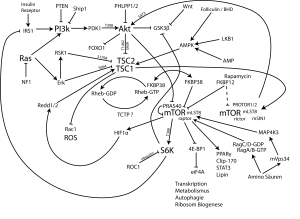
MTOR-pathway-ger Among the subtypes are those that respond to metabolic inhibitors such as the PI3K and mTOR directed drugs. Another subset may respond to drugs that target epidermal growth factor. There are basal-types that may be somewhat more refractory to therapy, while a subset may have biology related to the BRCA mutants, characterized by DNA repair deficiencies and exquisite sensitivity to Cisplatin-based therapies. Finally, a last group is associated with androgen signaling and may respond to drugs that target the androgen receptor.

Some years ago, we used the EVA-PCD platform to study refractory patients with breast cancer and identified exquisite sensitivity to the combination of Cisplatin plus Gemcitabine in this patient group. We published our observations in the Journal of Clinical Oncology and the combination of Cisplatin or Carboplatin plus Gemcitabine has become an established part of the armamentarium in these patients.

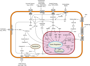
The I-SPY-2 trial has now used genomic analyses confirming our observations for the role of platins in TNBC. This iSignal_transduction_pathways.svgn part reflects the DNA repair deficiency subtype associated with the BRCA-like biology. More recently, we have examined TNBC patients for their sensitivity to novel therapeutic interventions. Among them, the PI3K and mTOR inhibitors, as well as the glucose metabolism pathway inhibitors like Metformin. Additional classes of drugs that are revealing activity are the cyclin-dependent kinase inhibitors, some of which are moving forward through clinical trials.

One feature of triple negative breast cancer is avid uptake on PET scan. This reflects, in part, the proliferation rate of these tumors, but may also reflect metabolic changes associated with altered glucose metabolism. In this regard, the use of drugs that change mitochondrial function may be particularly active. Metformin, a member of the biguanide family influences mitochondrial metabolism at the level of AMP kinase. The activity of Metformin and related classes of drugs in triple negative breast cancer is a fertile area of investigation that we and others are pursuing.

When we examine the good response of many triple negative breast cancers to appropriately selected therapies, the potential for durable complete remissions and the distinctly different biology that TNBC represents, the question arises whether TNBC is actually a worse diagnosis, or simply a different entity that requires different thinking. We have been very impressed by the good outcome of some of our triple negative breast cancer patients and believe this a very fertile area for additional investigation

The Information Disconnect

I recently had an interesting conversation with a physician regarding her patient with an aggressive breast cancer.

A portion of tumor had been submitted to our laboratory for analysis and we identified activity for the alkylating agents and the Taxanes, but not for Doxorubicin. After our report was submitted to the treating physician she contacted me to discuss our findings, as well as the results from a genomic/proteomic laboratory that conducted a parallel analysis upon a portion of the patient’s tumor. The physician was kind enough to forward me their report. Their results recommended doxorubicin while ours did not. The treating physician asked for my input. Here, I thought, was a “teachable moment.”

Our discussion turned to the profound difference between analyte-based laboratory tests e.g. genomic and proteomic, and functional platforms like our own (EVA-PCD). Genomic, transcriptomic and proteomic platforms measure the presence or absence of genes, RNA or protein. Gene amplification, deletions or mutations and protein and phosphoprotein expressions are examined. These platforms dichotomize patients into those who do and those who do not express the given analyte, with cutoffs for gene copy number or intensity of staining.

These platforms have worked very well in diseases where there is a linear connection between the gene (or protein) and the disease state, e.g. BCR-ABL in CML for which imatinib has proven so effective. These tests have worked reasonably well in EGFR mutated and ALK gene rearranged in lung cancer, but even here response rates and response durations have been less dramatic. However, they have not worked very well at all for the vast majority of cancers that do not carry specific and well-characterized targets. These cancers reflect polygenic phenomena and are not defined by a single gene or protein expression.

Functional platforms look at cellular response to injury at the systems level and measure the end result of drug exposure to gauge the likelihood of a clinical response. Our focus on cell biology allows us to determine whether a drug or combination induces programmed cell death. After all, regardless of what gene elements are operational, it is the ultimate eradication of the cancer clone (its loss of viability) that results in clinical response.

As we reported in a recent paper in non-small cell lung cancer, patients who revealed the most sensitive ex-vivo profile to erlotinib (Tarceva) lived far longer than the general clinical experience for those patients who were selected for erlotinib by EGFr mutation analysis alone. Some of these patients are alive at 5, even 9 years since diagnosis.

We live in a technocracy where process has taken precedence over results. We are enamored with complex scientific technologies sometimes at the expense of simple answers. A metallurgist, familiar with every last detail of the alloys used in a Boeing 747 wouldn’t necessarily be your first choice for pilot. A skilled pathologist, intimately familiar with the most detailed intricacies of human diagnostics would not likely be your preferred surgeon for cardiac bypass.

Cancer diagnosis and cancer treatment are two distinctly different disciplines. While we use the ER (estrogen receptor) status in breast cancer to select treatment, few oncologists would select Tamoxifen for their NSCLC patients even though many NSCLC patients express ER in their tissue. ER + NSCLC does not respond to tamoxifen and V600E BRAF mutated (+) colon cancer patients do not respond to vemurafenib, the very drug that works so well in BRAF V600E (+) melanoma.

Cancer is contextual and responses are not solely predicated upon the presence or absence of a gene element alone. We must use a broader brush when we paint the likeness of our patients in the laboratory, one that encompasses the vicissitudes of human biology in all of its complexities.

Where I took issue with the report, however, was its “evidence-based” moniker.  The evidentiary manuscripts cited to support the drug recommendations, with titles like “Overexpression of COX-2 in celecoxib-resistant breast cancer cell lines” provided little evidence that a (+) COX-2 finding by IHC on this patient’s biopsy specimen would offer any real hope of response. It seemed that with all of the really interesting science going on here, no one had taken the time to do the hard work to figure out whether any of these observations had a basis in reality. The failure of ERCC1 expression in lung cancer to correlate with response and survival or the Duke University debacle with gene profiling in NSCLC are just the most recent examples of how “lovely theories can be spoiled by a little fact.”

As we and our colleagues in cell profiling have actually taken the time to correlate predictions with clinical outcomes we have shown a 2.04 fold higher objective response rate (p 0.001) and significantly improved 1-year survival (p=0.02). (Apfel, C. et al Proc ASCO, 2013). To the contrary, it is of interest to examine the comparatively scant published literature on genomic and IHC profiling for drug selection under similar circumstances. While one group reported an underwhelming objective response rate of 10 percent in their study, (Von Hoff, J Clin Oncol 2010) a more recent study is even more illuminating. A Spanish group used genomic profiling in 254 colon cancer patients to select candidates for gene-targeted agents (KRAS/BRAF/PI3K/PTEN/MET) and provided therapy for 82. They reported a significantly shorter time to progression for targeted treatments compared with conventional therapies 7.9 vs 16.3 week (P<0.001) and an overall objective response rate of 1.2 percent, yes that’s 1.2% (1/82).

Human tumor biology is many things, but simple is not one of them. Reductionist thinking is not providing the insights that our patients desperately need. While we await the arrival of a perfect test for the prediction of response to cancer therapy, perhaps we as physicians and our patients should use a good one, one that works.

The 2013 Nobel Prize for Medicine and Physiology

2013 Nobel Prize artOn October 7, 2013, in Stockholm, Sweden, the Nobel Committee announced the winners of the Nobel Prize for Medicine and Physiology – two Americans and one German, all now located at institutions in the US. The discovery for which these three investigators share the prize involves their work over three decades studying the transport, packaging and trafficking of cellular proteins.

All cells must communicate and maintain their identity. To do so cells have developed intricate systems whereby neurotransmitters, proteins, hormones, and other species are encapsulated in small vesicles. These vesicles may be utilized to extrude materials into the extracellular domain or may store materials within the cell for later use. Working in model systems including yeast cells, these investigators showed the intricacy of cellular physiology associated with micro-vesicular function.

What makes these investigators’ work so interesting is that it is principally the study of cellular physiology, or what we call cell biology. While many breakthroughs and observations today reflect discoveries at the level of DNA, RNA and the genome, these investigators have pioneered protein kinetics and physiology. What is so exciting about this Nobel Prize is that it returns attention to the intricacies of cellular function at the level of phenotype. Protein biology represents the final common pathway from blueprint (DNA) to function. While genes that are detected within the nucleus (the purview of genomic analyses and many recent Nobel prizes) may or may not ultimately be expressed, depending upon splice variants, DNA methylation, histone acetylation, small interfering RNAs and non-coding DNAs among other phenomena, functional proteins are the active end-product and do very much exist.

We now recognize that cellular signaling, misfolded protein response, autophagy and apoptotic responses are tightly bound together. Among the most toxic phenomena for a cell is the misfolded protein signal, a signal that occurs far from the gene. This represents the target of the newest classes of drugs known as proteasome inhibitors and heat shock protein inhibitors, which function within the cytoplasm, not the nucleus.

It is exciting to imagine a day when physiologist, biochemist, enzymologist, physical chemist, and protein chemist regain their position as leaders in cancer research.

N.B: It should not go unmentioned that the EVA-PCD® assay offered by Rational Therapeutics is based on cellular function.